PDH Complex, Fatty Acid Beta-Oxidation, and Introduction to the Krebs Cycle
Date
2026-08-17
Source transcript
transcripts/en-BIS-103_ 2026-08-17 12_07.txt
Lecturer
Dr. Hill
Subject Area
BIS 103 — Biochemistry
Lecture Overview
This lecture continues the discussion of the pyruvate dehydrogenase (PDH) complex, beginning with the structure and properties of FAD and the detailed mechanism of E1, E2, and E3. The lecture then transitions to fatty acid catabolism: activation of fatty acids by fatty acyl-CoA synthetase, transport into the mitochondrial matrix via the carnitine shuttle, and breakdown by the four enzymes of beta-oxidation. The final portion introduces the Krebs cycle, covering the first five steps from acetyl-CoA through succinate, including a detailed comparison of the alpha-ketoglutarate dehydrogenase complex to the PDH complex.
Key Concepts and Definitions
Thioester: A compound with a sulfur connected to a carbonyl group; higher energy than an oxygen ester due to less resonance stabilization from sulfur (sulfur is not very electronegative).
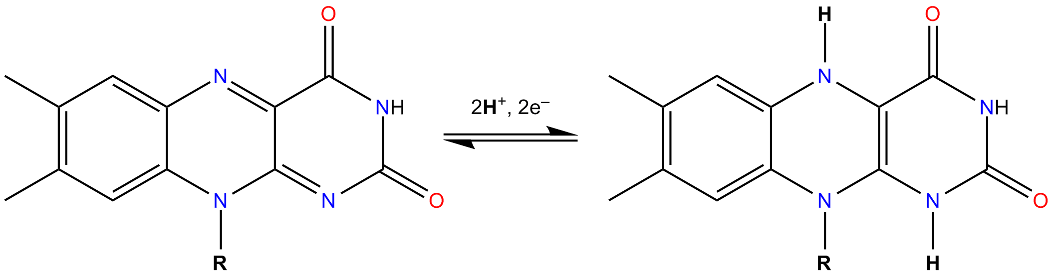
FAD (flavin adenine dinucleotide): A coenzyme whose business end is the isoalloxazine ring (a tricyclic ring); can accept one or two electrons; does not have a single published redox value because its potential depends on the protein it is bound to.
FMN (flavin mononucleotide): A portion of FAD lacking the adenine nucleotide; shares the isoalloxazine ring and the same electron-handling properties.
Semiquinone (FADH): The partially reduced, one-electron form of FAD.
Lipoic acid: A cofactor on the E2 tether containing a disulfide bond; must have the disulfide bond to be active; can also carry an acetyl group as a thioester.
Tether: A long flexible structure on E2 (lysine residue plus lipoic acid) that swings between enzyme active sites in the PDH complex.
Fatty acyl-CoA synthetase: The enzyme that activates fatty acids by linking them to CoA as a thioester, using ATP (which yields AMP + pyrophosphate).
Synthetase vs. synthase: Synthetase (-thetase) uses a nucleoside triphosphate (usually ATP) as energy source; synthase (-thase) does not.
Carnitine: A small, net-zero-charge molecule derived from lysine that carries fatty acids across the mitochondrial inner membrane.
CAT (carnitine acyltransferase): Enzyme transferring fatty acids between CoA and carnitine; CAT1 on the outer membrane, CAT2 on the matrix side.
Beta-oxidation: The primary pathway for breaking down fatty acids by sequentially removing two-carbon units as acetyl-CoA, using four enzymes per cycle.
Krebs cycle (citric acid cycle / TCA cycle): A circular metabolic pathway in the mitochondrial matrix that oxidizes acetyl-CoA, producing NADH, CO2, and GTP.
Citrate synthase (condensing enzyme): Catalyzes the first step of the Krebs cycle, condensing acetyl-CoA (2C) onto oxaloacetate (4C) to form citrate (6C).
Aconitase: Converts citrate to isocitrate; requires a 4-iron, 4-sulfur complex as cofactor; a moonlighting protein that also senses cellular iron status.
Moonlighting protein: A protein that performs two different functions; aconitase is the example given.
Succinyl-CoA: A four-carbon thioester intermediate in the Krebs cycle; high energy.
Alpha-ketoglutarate (alpha-KG): A five-carbon Krebs cycle intermediate.
Chronological Lecture Notes
PDH complex
1. Exam logistics
The second midterm is Wednesday.
Last names A through N in the regular room; remaining students in room 1215 TLC.
Tests distributed about five minutes after 12; students have 50 minutes.
Bring a pen or pencil and a calculator.
No questions answered during the exam because the 50-minute window is too short and interruptions are disruptive.
Questions are straightforward with no tricks intended.
Equations and constants will be provided; if something is not listed, it is not needed.
Material currently being covered is second midterm content.
2. PDH complex and thioesters (continued from previous lecture)
The pyruvate dehydrogenase (PDH) complex is a very large structure that floats in the matrix of the mitochondria.
Thioesters (sulfur connected to a carbonyl group) are at a much higher energy level than oxygen esters.
Oxygen provides more resonance stabilization than sulfur because sulfur is not very electronegative.
Cells use thioesters to release energy via large negative delta G values.
3. FAD and FMN: structure and properties
FAD (flavin adenine dinucleotide) is introduced for the first time in this course.
The business end of FAD is the isoalloxazine ring, a tricyclic ring structure. Students should be able to recognize the structure and know the ring name, but will never need to draw it.
FMN (flavin mononucleotide) is a smaller version of FAD that also contains the isoalloxazine ring. Whatever is said about FAD applies to FMN.
FAD does not have a single published redox value. Its redox potential depends on the protein it is bound to. FAD is always bound by weak bonds to the active site, and the amino acid residues affect its electron properties.
This variable redox potential is particularly important in the electron transport chain (ETC).
NADH is free (dissociates from enzymes) and typically has a much higher redox potential than FAD. FAD has two advantages over NADH:
FAD can adopt a spectrum of different redox values depending on its protein environment, making it very versatile.
FAD can accept one electron or two electrons, whereas NADH can only accept two electrons as a hydride (H minus).
Electrons are taken on by FAD as H dot (an electron accompanied by a proton).
FAD and FMN act as a "gear shift": most of the electron transport chain handles only one electron at a time, so when NADH delivers two electrons, FAD or FMN (bound to a protein) can release them safely one at a time.
A single unpaired electron is normally a dangerous free radical, but in FAD the electron is spread by resonance over the entire isoalloxazine ring, making it stable.
Reference figure · external sourceRedox equilibrium between FAD and FADH2, showing the isoalloxazine ring that accepts electrons in the flavin coenzymes discussed in this section. Source: Wikimedia Commons · DMacks · CC BY-SA 3.0
4. PDH complex: structural model
The PDH complex resembles a large soccer ball. The model shown in Leninger is cut in half to reveal the interior.
The term "model" means the full structure is not definitively known; this is the current best guess.
The complex contains many copies of E1, E2, and E3.
E2 has tethers: long structures that carry intermediates from one enzyme active site to another.
The model is based on bovine (cow) research.
X-ray crystallography cannot be performed on the intact complex because it is approximately 10 million molecular weight; crystallizing such a massive assembly in perfect alignment is not feasible with current technology.
E1, E2, and E3 have been individually isolated and crystallized, but their conformations change when they interact within the complex.
5. PDH complex: detailed mechanism of E1, E2, and E3
E1 (yellow): Active site contains thiamine pyrophosphate (TPP), a carbanion and strong nucleophile. TPP attacks pyruvate at the carbonyl carbon, releasing CO2 (a source of exhaled CO2 in animals). The remaining two carbons are carried covalently by TPP.
E2 (green): Has an active site and a tether. The tether consists of a lysine residue (four carbons long) with lipoic acid attached at the amino end. Lipoic acid contains a disulfide bond.
The tether swings into E1's active site. A hydride from the two-carbon intermediate reduces one sulfur of the disulfide. The other sulfur carries the two-carbon unit as a thioester — a high-energy molecule.
The tether then carries this thioester to E2's active site where CoA (in reduced form) enters. The enzyme transfers the two-carbon thioester from lipoic acid's sulfur to CoA's sulfur, forming acetyl-CoA. This transfer is energetically inexpensive because one thioester is exchanged for another of essentially the same energy.
Acetyl-CoA diffuses to the Krebs cycle, which is also in the mitochondrial matrix.
E3 (pink): Has FAD bound to it. After the tether has delivered its cargo, both sulfurs on lipoic acid are reduced. E3's FAD takes the two electrons and protons from the reduced sulfurs to regenerate the disulfide bond. NAD then arrives at E3's active site and receives the electrons as a hydride, forming NADH plus a proton.
NADH goes to complex I of the electron transport chain.
The tether is now regenerated (disulfide bond restored) and ready for the next pyruvate.
Exam emphasis: The lecturer stressed knowing this mechanism very well, because life reuses this type of multi-enzyme complex scenario, particularly in the Krebs cycle.
A student asked why FAD is used at this step. The lecturer responded that "why" questions are difficult to answer definitively; research shows this is what the enzyme does. The key point is that the NADH produced here, along with all other NADHs, goes to complex I of the electron transport chain.
6. Lipoic acid structure and tether variations across species
The tether on E2 consists of part of the protein peptide chain, the lysine residue, and lipoic acid.
Students should be able to recognize lipoic acid if shown its structure.
Active lipoic acid requires a disulfide bond; it can also be fully reduced or carrying an acetyl group.
E2 has three domains: (1) the enzyme active site catalyzing the reaction, (2) a domain interacting with neighboring E2 molecules, and (3) a domain carrying the tether.
Tether numbers vary by species:
Yeast: 1 tether per E2
Mammals (e.g., cows): 2 tethers per E2; cows have 60 copies of E2 in their PDH complex, yielding 120 tethers total
E. coli: 3 tethers per E2, but fewer copies of other enzymes
Evolution has tweaked the tether arrangement to best fit each organism.
7. Disulfide exchange and tether dynamics within the PDH complex
Inside the PDH complex, the many tethers can move and collide with each other.
Disulfide exchange reactions: An SH group on one tether attacks the disulfide bond on another tether, transferring the disulfide bond. This is not enzyme-catalyzed; it results from the very high local concentration (molarity) of tethers confined within the small interior volume of the complex.
Acetyl group transfer: An SH group can also attack the carbonyl carbon of a thioester on another tether (partial positive charge from oxygen drawing electron density away), transferring the acetyl group between tethers.
These exchange reactions occur constantly and in all directions.
8. PDH complex efficiency
The lecturer demonstrated the complex's efficiency using students as E2 molecules, one student as E3, and the lecturer as E1.
E1 rapidly loads an acetyl group onto a nearby E2 tether. E2 tethers bump into each other, transferring intermediates among themselves. E3 removes reduced products.
The PDH complex takes in pyruvate and produces acetyl-CoA and NADH with remarkable efficiency.
The same type of mechanism operates in a related complex in the Krebs cycle and in some other enzymes.
Fatty acid activation and beta-oxidation
9. Fatty acid breakdown: overview and activation
Shifting from PDH to the breakdown of fatty acids.
Context: glucose undergoes glycolysis in the cytosol to pyruvate, which enters the mitochondria for the PDH reaction. Fatty acids follow a separate route.
Fatty acids must be activated before enzymes can break them down: the fatty acid is covalently linked to coenzyme A (CoA) as a thioester.
Activation occurs on the surface of the endoplasmic reticulum (ER), facing the cytosol.
Once activated, fatty acids enter the mitochondrial matrix and are broken down primarily by beta-oxidation (the main pathway; alpha- and gamma-oxidation also exist but are not the focus).
Long-chain fatty acids (e.g., 16 or 18 carbons) are chopped into two-carbon units, generating many molecules of acetyl-CoA.
10. Fatty acid activation reaction
Using palmitate (16 carbons, ionized) as an example. All five fatty acids from the course must be activated the same way.
The enzyme fatty acyl-CoA synthetase links the carbonyl carbon of the fatty acid to the sulfur of CoA, forming a thioester (e.g., palmitoyl-CoA).
Three substrates: the fatty acid, CoA, and ATP.
The suffix "-thetase" (synthetase) indicates a nucleoside triphosphate (usually ATP) is used as the energy source; the suffix "-thase" (synthase) indicates the energy source is not a nucleoside triphosphate.
ATP is broken down differently here: it yields AMP (adenosine monophosphate) plus pyrophosphate (PPi, two phosphates connected together), rather than ADP plus inorganic phosphate.
This mode of ATP hydrolysis releases more energy: delta G' = -9.9 kcal/mol, compared to -7.7 kcal/mol for the standard ATP to ADP + Pi hydrolysis. (Values do not need to be memorized; they will always be provided.)
Historical note: Professor Paul Stumpf, one of the founders of the UC Davis biochemistry department, referred to this as the "CoA track."
Exam emphasis: The lecturer expects students to be able to draw the activation reaction on the second midterm or final.
11. Pyrophosphate hydrolysis drives activation
A separate, ubiquitous enzyme called pyrophosphatase hydrolyzes pyrophosphate (PPi) into two inorganic phosphates using water.
The pyrophosphatase attacks the phosphorus (partial positive charge) with water acting as a nucleophile (enhanced by the enzyme's active site).
Removing pyrophosphate (a product of the activation reaction) pulls the original reaction further to the right.
Delta G' of pyrophosphate hydrolysis: -4.2 kcal/mol.
Overall delta G': (-9.9) + (-4.2) = -14.1 kcal/mol, almost equivalent to 2 ATP.
In effect, activating a fatty acid costs the equivalent of 1 ATP (since 2 high-energy phosphate bonds are consumed).
This cost is the same regardless of fatty acid chain length: one ATP equivalent activates the fatty acid onto the CoA track.
Only fatty acids linked to CoA can enter the mitochondria and be broken down.
CoA functions like a "zip code": it often signals that a molecule is headed to the mitochondria for destruction.
12. Carnitine and the carnitine shuttle
Carnitine is the carrier that transports fatty acids across the mitochondrial inner membrane.
Properties making carnitine suited for this role:
Small molecular weight — diffuses faster.
No net charge — permanent +1 on nitrogen, permanent -1 on carboxylate group; net charge is always zero. This prevents charge buildup across the membrane.
A hydroxyl group serves as the site where the fatty acid is attached (as an oxygen ester).
Carnitine is derived from the amino acid lysine: the epsilon amino group of lysine becomes the nitrogen, which is methylated three times.
The enzyme carnitine acyltransferase (CAT) transfers the fatty acid from the CoA thioester to carnitine's hydroxyl group, forming an oxygen ester. The thioester energy drives this transfer. Free CoA is released.
The transport protein in the inner membrane only recognizes two molecules: acylcarnitine (carnitine bound to a fatty acid) and free carnitine.
13. Fatty acid transport across mitochondrial membranes
Outer membrane (OM): Has large pores allowing anything of 5,000 molecular weight or smaller to pass through freely.
CAT1 (isomer 1): Attached to the outer membrane. Whether its active site faces the cytosol or the intermembrane space is unknown, but it does not matter because substrates can access it either way. CAT1 converts fatty acyl-CoA + carnitine into acylcarnitine.
Acylcarnitine/carnitine transport protein: An integral protein spanning the entire inner membrane. It allows acylcarnitine to enter the matrix only if a free carnitine exits simultaneously. This is an antiport system driven by facilitated diffusion (entropy/delta S): molecules move from high concentration to low concentration, one-to-one.
CAT2 (isomer 2): Located on the matrix side. Has a slightly different amino acid sequence from CAT1 but catalyzes the same reaction in reverse: it transfers the fatty acid from carnitine back onto CoA, regenerating the fatty acyl-CoA thioester in the matrix. Free carnitine is released for export.
Purpose of the shuttle: The carnitine system keeps the CoA pool inside the mitochondrial matrix separate from the CoA pool in the cytosol. CoA is needed for many different biochemical reactions in both compartments; the mitochondria do not share their CoA with the cytosol. Cytosolic CoA is used for building phospholipids and other membrane components.
14. Beta-oxidation: the four enzymes
Four enzymes in the mitochondrial matrix break palmitoyl-CoA down into acetyl-CoA units.
The Krebs cycle only accepts acetyl-CoA, so long-chain fatty acids must be cut into two-carbon units.
Carbon numbering: C1 is the carbonyl carbon; C2 is the alpha carbon (adjacent to the carbonyl); C3 is the beta carbon.
Creates a trans double bond between the alpha and beta carbons (C2 and C3). Hydrogens are on opposite sides (never the same side). Students must draw this as a trans double bond.
FAD is the coenzyme. It never leaves the enzyme and is never consumed. Its redox potential is determined by the enzyme.
Electrons are passed through the enzyme and other proteins directly into the electron transport chain (FAD does not diffuse away as FADH2).
Free CoA attacks the beta-carbonyl carbon (which bears a partial positive charge because the oxygen draws electron density away). The enzyme positions CoA's sulfur precisely next to this carbon, never the other carbonyl.
Cuts the bond between the alpha and beta carbons.
Products: acetyl-CoA (2 carbons) and a fatty acyl-CoA that is two carbons shorter, still carried on CoA.
Acetyl-CoA goes to the Krebs cycle.
A student asked about enzymes recognizing different double bond positions. The lecturer noted that in reality there are different forms of these enzymes for different substrates, but for this course, each of the four enzymes is treated as handling everything.
15. Beta-oxidation: cycle counting and products from palmitate
All four enzymes acting in sequence constitutes one cycle of beta-oxidation, releasing one acetyl-CoA per cycle.
The shortened fatty acyl-CoA re-enters the same four-enzyme sequence.
When only four carbons remain on CoA, the thiolase reaction produces two acetyl-CoAs (not one) and there is nothing left to process.
For palmitate (16 carbons): 7 cycles of beta-oxidation occur (the first cycle plus 6 more), producing a total of 8 acetyl-CoAs.
Each cycle produces 1 NADH and 1 FADH2, so 7 cycles yield 7 NADHs and 7 FADH2s.
Comparison: one molecule of palmitate produces 8 acetyl-CoAs; one molecule of glucose produces only 2 acetyl-CoAs. Palmitate provides far more energy.
Regardless of origin, all acetyl-CoAs feed into the Krebs cycle.
16. Unsaturated fatty acids and beta-oxidation
Oleic acid, linoleic acid, and alpha-linolenic acid present two problems for beta-oxidation:
They contain one or more cis double bonds, but the first enzyme of beta-oxidation requires a trans double bond.
As carbons are removed, the pre-existing double bonds are not necessarily positioned between the alpha and beta carbons.
Two additional enzymes work together to solve both problems (an isomerase and a reductase), but their detailed pathways are not covered in this course.
All unsaturated fatty acids are fully utilized by the cell.
Krebs cycle
17. Introduction to the Krebs cycle
The Krebs cycle occurs in the matrix of the mitochondria.
Three equivalent names: Krebs cycle (honoring the scientist who did most of the isolation work), citric acid cycle (citrate is an intermediate), and TCA cycle (citrate is a tricarboxylic acid). Any name is acceptable on exams.
The cycle begins with acetyl-CoA (two carbons, high energy).
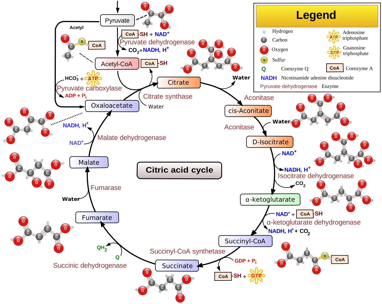
Reference figure · external sourceOverview of the citric acid cycle, showing the circular pathway and intermediates discussed from step 1 (citrate synthase) through step 5 (succinyl-CoA synthetase) in this lecture. Source: Wikimedia Commons · Narayanese, WikiUserPedia, YassineMrabet, TotoBaggins · CC BY-SA 3.0
18. Acetyl-CoA chemistry
The carbonyl carbon of acetyl-CoA has a partial positive charge (mostly from oxygen, slightly from sulfur), making it a target for nucleophilic attack.
The alpha hydrogen (on the methyl group adjacent to the carbonyl) is slightly acidic. Enzymes facilitate removal of this hydrogen, allowing the alpha carbon to act as a nucleophile.
Acetyl-CoA therefore has both a nucleophilic site (alpha carbon) and an electrophilic site (carbonyl carbon). Both features are used in the first reaction of the Krebs cycle.
The alpha carbon of acetyl-CoA attacks a carbon on oxaloacetate (OAA).
The enzyme is called citrate synthase (suffix -thase: energy source is not a nucleoside triphosphate). The energy is provided by the thioester bond, which has more energy than ATP.
Water enters the same active site and releases CoA, freeing it for other mitochondrial reactions.
Common name: condensing enzyme (condenses two carbons onto a four-carbon acceptor).
The two carbons brought in by acetyl-CoA will eventually leave the cycle as CO2 in later steps.
20. Krebs cycle step 2: Aconitase
Citrate --[Aconitase]--> Isocitrate
Aconitase moves a hydroxyl group from one carbon to the adjacent carbon below it.
Mechanism: the enzyme removes a water molecule and then puts a different water molecule on at the new position (it is easier to remove and add water than to move an OH group directly, because water has no charge).
Leninger shows cis-aconitate as an intermediate of this reaction, but this is just a mechanistic intermediate. Students are not to include cis-aconitate when drawing the Krebs cycle; the step is simply citrate to isocitrate.
Cofactor: Aconitase requires a 4-iron, 4-sulfur (4Fe-4S) complex at its active site. Without it, the enzyme does not function.
Three of the four irons are bound by cysteine residues and have four bonds to sulfurs each.
The fourth iron has only three sulfur bonds and is directly involved in chemistry with the substrates (citrate and water).
The nomenclature "4-iron, 4-sulfur" does not count the sulfurs from cysteine side chains.
21. Aconitase as a moonlighting protein
When substrates (citrate, water) are absent, the fourth iron is held less strongly and can leave the complex. When it leaves, the remaining complex does not function.
Loss of this iron triggers a separate set of reactions that sense insufficient iron in the cell.
Aconitase is therefore a moonlighting protein — a protein with two distinct jobs:
Krebs cycle enzyme (catalyzing citrate to isocitrate).
Cellular iron sensor (triggering iron-deficiency responses when the fourth iron is lost).
This is a multi-enzyme complex (E1, E2, E3 in many copies, forming a smaller soccer ball), structurally and mechanistically similar to the PDH complex.
Another CO2 is released. At this point, both carbons originally brought in by acetyl-CoA have been lost as CO2.
The remaining cycle must regenerate oxaloacetate so the circular pathway can continue indefinitely. Only small amounts of intermediates are needed.
Some Krebs cycle reactions are irreversible (very negative delta G), partly because CO2, a product, escapes, pulling reactions forward.
Three separate NADHs are produced in the cycle (from steps 3, 4, and later step 8); all go to complex I of the electron transport chain.
The product succinyl-CoA is a four-carbon thioester (high energy, more energy than ATP).
Comparison of alpha-KG dehydrogenase complex to PDH complex:
Feature
PDH complex
Alpha-KG dehydrogenase complex
E1 substrate
Pyruvate (3C)
Alpha-ketoglutarate (5C)
E1 cofactor
TPP
TPP (same cofactor)
Carbons carried by TPP after CO2 loss
2
4
E2 tether
Lipoic acid (same)
Lipoic acid (same)
E2 thioester product
Acetyl-CoA (2C)
Succinyl-CoA (4C)
E3
FAD, produces NADH + H+
Identical enzyme to PDH E3
E1 amino acid sequences differ because the two complexes recognize different substrates, but the end of alpha-KG structurally resembles pyruvate.
E2 is similar but not identical (different products, slightly different active site).
E3 is the same protein in both complexes: it removes electrons and protons from the same lipoic acid to produce NADH + H+.
24. Krebs cycle step 5: Succinyl-CoA synthetase
Succinyl-CoA + GDP + Pi --[Succinyl-CoA synthetase]--> Succinate + GTP + CoA
The energy stored in the succinyl-CoA thioester is transferred to GDP to make GTP.
Three substrates: succinyl-CoA, GDP (guanosine diphosphate), and inorganic phosphate.
GTP has the same amount of energy as ATP.
Some organisms make ATP at this step instead of GTP, but most make GTP. The course uses GTP.
The lecturer speculated that this may help supply GTP for GPCR signaling (G-protein-coupled receptors), but noted this is not confirmed.
Succinate is four carbons long. A line of symmetry between the top two and bottom two carbons makes them mirror images. Because of this symmetry, the two carbons originally from acetyl-CoA can no longer be distinguished from the other two. This is why the "pink" carbon labeling from acetyl-CoA disappears at the succinate stage.
Study Review Questions
What are the three oxidation states of FAD, and what is unique about the semiquinone form?
Why does FAD lack a single published redox potential, and how does this property make FAD versatile in the electron transport chain?
Describe the roles of E1, E2 (including the tether), and E3 in the PDH complex mechanism, including the cofactor each uses.
Why is the transfer of the acetyl group from lipoic acid to CoA energetically inexpensive in the PDH complex?
In the activation of fatty acids, why does the cell use pyrophosphatase in conjunction with fatty acyl-CoA synthetase, and what is the overall energy cost?
What properties of carnitine make it well suited for transporting fatty acids across the mitochondrial inner membrane?
Explain why the carnitine shuttle system is used rather than directly transporting fatty acyl-CoA into the matrix.
List the four enzymes of beta-oxidation in order, name the cofactor for each (if any), and state the chemical change each performs.
How many acetyl-CoAs, NADHs, and FADH2s are produced from the complete beta-oxidation of one molecule of palmitate (16 carbons)?
What makes aconitase a "moonlighting protein," and what are its two distinct functions?