Lecture Overview
The lecture first develops microcalorimetry as a way to quantify heat flow from living cells and then shows how a modified experiment can also yield carbon dioxide production and oxygen consumption rates. It next introduces intermediary metabolism, pathway shapes, and the use of reaction coupling to make glutamine from glutamate and ammonium. The final portion begins glycolysis with glucose transport, hexokinase, magnesium, glucose-triggered enzyme closure, and the two-ATP investment that precedes the aldolase split. The lecturer was aiming to close the first-midterm material at the end of Monday and asked students to learn the pathway over the four-day weekend.
Course Boundary and Study Preparation
The lecturer was aiming to finish the first-midterm material by the end of Monday, draw a line there, and then begin second-midterm material.
- This was presented as a plan rather than a guarantee.
- With the first exam approaching, students were asked to introduce themselves and identify possible study partners.
Measuring Heat Flow from Living Cells
Microcalorimetry measures the very small flow of heat from cells that remain alive.
- This differs from calorimetry in freshman chemistry, where material is burned to completion and the total heat released is measured.
- The instrument was initially designed to measure heat rate. With modifications, it can also provide rates of
CO2production andO2consumption. - The lecturer described eukaryotic-cell
CO2as coming mostly from mitochondria and mitochondria as the principal organelles using inhaled oxygen.
The free-energy frame
The Gibbs free-energy equation was used to locate what the instrument measures:
ΔGis energy available to do work.ΔHis the heat or enthalpy change.Tis temperature in Kelvin.ΔSmeasures entropy or disorder.- The lecturer described heat and
TΔSordering as two contributions to free energy; the microcalorimeter measures onlyΔH.
Exam emphasis: The equation will be supplied on exams. Students are expected to understand its concepts, while calculations with
ΔGwere deferred until later in the course.
Sensitivity and steady state
A 0.7-watt light-emitting diode (LED) night-light illustrated the instrument's sensitivity.
- A watt is a joule per second, so it is a rate.
- The light was treated as an open system at steady state: energy enters and leaves, mostly as light and partly as heat. Its heat output was assumed to be proportional to its light output.
- The lecturer dimmed the light by one-seventh and then by successive factors of 10, describing a 70-fold reduction before continuing downward.
- This analogy approached the roughly one-microwatt range that the instrument can detect.
- Steady state was emphasized as important for living cells and enzyme kinetics.
The Modified Microcalorimeter
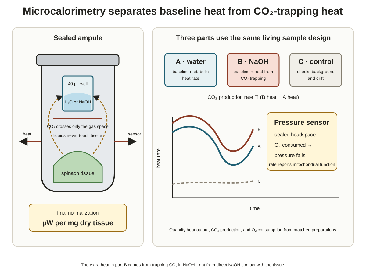
A sealed metal ampule transfers cellular heat to sensors while a separate internal well and pressure line add chemical and gas information.
- Professor Richard Criddle, the retired campus microcalorimetry expert who taught the lecturer, routinely used the modifications described here.
- The machined metal-alloy ampule is about a thumb segment in size, has a volume of about “one mil” in the lecturer's wording, and transfers heat well. Larger instruments can hold more tissue, but the lecturer did not specify the problems they introduce.
- A small metal well is welded inside the ampule and initially receives about 40 microliters of water.
- Plant leaves, red blood cells, bacteria, or other material can occupy the ampule, but the sample must not touch the liquid in the well.
- A tightly screwed lid contains a tiny tube connected by thin fiber-optic tubing to a pressure sensor. Thin tubing limits heat conducted into the ampule from outside.
- The welded well and pressure sensor are optional modifications, not requirements of every microcalorimetry experiment.
Sample positions and heat detection
- Enough cells must be loaded for the signal to exceed background. About 10 leaves was one example, not a fixed requirement.
- A snug opening surrounds the ampule with heat sensors in three dimensions. Heat crosses the metal wall, and the sensors convert it to an electrical signal for the computer.
- A typical instrument has four openings: one unchanged, empty reference ampule and three experimental positions.
- Example experimental positions included 10 normal leaves, 10 pesticide-treated leaves, and either a duplicate or a third condition. The pesticide comparison was connected to a homework example.
- Each experimental ampule has its own pressure sensor and sends a separate channel to the computer.
- Individual notched lids and a second overall lid shield the extremely sensitive heat sensors from room heat.
What the plot reports
- The heat-flow y-axis is microwatts, or joules per second, per milligram dry weight.
- The x-axis is time in minutes, and a typical complete run lasts about 90 minutes.
- Dry weight corrects for unequal tissue mass when cells cannot be counted directly, unlike bacteria or red blood cells.
The Three-Part Heat Experiment
The difference between a water baseline and an excess-sodium-hydroxide plateau reveals the heat associated with trapping all produced CO2, while a return to water checks for damage.
- Part A — establish the living-cell baseline. Opening the instrument causes an off-scale room-heat transient that is ignored while the instrument recovers. The later plateau is cellular heat flow. A value around 80 was only an illustrative healthy signal above the approximately one-microwatt detection floor; a plateau at zero means the cells are dead and the experiment must restart. The example ampule held about 10 leaves and 40 microliters of water in the separate well.
- Part B — trap
CO2. After an illustrative 30 minutes at steady state, remove the 40 microliters of water and replace it with 40 microliters of 0.4 M sodium hydroxide (NaOH) in large excess, without letting it touch the tissue. Resealing three ampules may take 5–10 minutes and produces another off-scale transient. Part B usually reaches a higher plateau. The lecturer identified this as the Part B used in Homework 1, probably Question 2, but was unsure of the exact question number.
The lecturer corrected “distance” to difference when comparing the extrapolated Part-A plateau with Part B. That difference is proportional to the heat-rate change.
Why excess sodium hydroxide raises Part B
- Cells produce
CO2, and excess hydroxide is used so that all of it reacts rather than only a fraction. - Hydroxide attacks
CO2, first forming bicarbonate; additional hydroxide removes its proton to form carbonate. - The reaction releases heat. Because its heats of reaction are known, the measured heat can be converted to moles, nanomoles, or picomoles of
CO2produced per unit time, such as per minute.
- The two oxygens withdraw electron density from the carbon in
CO2, giving that carbon partial positive character and making it a target for a nucleophile. - Some reactions are endothermic, but this trapping reaction is used because it is exothermic.
CO2is nonpolar, leaves cells, moves rapidly through the sealed gas space, and is chemically trapped when it contacts theNaOHsurface.- If Part B equals Part A, no detectable
CO2is being produced. For plant cells, the lecturer said this would be bad and would probably mean the mitochondria were not working. - The known reaction heat is treated as accurately established. Excess base drives the sequence fully to carbonate, so the contributing heats can be added or looked up.
Part C restores the control condition
- Part C — restore water and test for damage. After about another 30 minutes to assure steady state, remove the
NaOHand restore 40 microliters of water. A meticulous cleanup may use a little hydrochloric acid (HCl), remove it, rinse once or twice with water, and then add the final water. After the transient, the plateau should return approximately to Part A.
- Part C tests whether something harmful happened during Part B. Accidental
NaOHcontact could destroy cells and release heat unrelated toCO2. - Part A contains water because most of a 0.4 M
NaOHsolution is water. This controls for the possibility that water vapor itself changes metabolism. - The lecturer announced a second reason for the water but did not clearly complete it, so no second reason is supplied here.
Normalization, Oxygen Consumption, and Applications
The completed experiment combines normalized heat flow, trapped-CO2 heat, and the Part-B pressure decrease to produce three numerical rates.
Dry-weight normalization
After the 90-minute run:
- Remove each tissue sample into its own container.
- Heat the samples at 90 degrees in a vacuum oven to remove water.
- Leave them overnight, then weigh each dried sample on a high-quality balance.
The vacuum removes oxygen so the tissue is not oxidized or burned and thereby made artificially lighter.
Isolating oxygen in Part B
- Pressure is recorded alongside heat for each experimental ampule.
- In Part A, nitrogen, oxygen,
CO2, and other gases contribute collectively, so a pressure change cannot be assigned to one gas. - In Part B,
CO2is chemically removed and nitrogen is inert. The falling pressure is therefore assigned to oxygen consumption, mainly by mitochondria. - Parts A and C contain too many changing gases to isolate a particular gas; only Part B provides this specificity.
- The Part-B decrease can be converted, for example, to picomoles of oxygen consumed per minute with
PV = nRT.
Exam emphasis: Students will not be required to use
PV = nRTbecause other material takes priority.
The three reported outputs are:
| Output | Basis of measurement |
|---|---|
| Heat rate per milligram dry weight | Cellular heat transferred through the ampule wall |
CO2 production rate |
Extra heat from complete trapping by excess NaOH |
O2 consumption rate |
Pressure decrease isolated during Part B |
- These numerical outputs are not provided by a DNA microarray or a 2-D-IEF-SDS-PAGE gel.
- In one application, small tree-tissue samples could identify high-metabolism, high-growth varieties in about 2–3 days rather than waiting 2–3 years to judge height qualitatively. The lecturer said these measurements can predict outcomes 10 years later.
- Cancer cells were another possible application.
- Student question: How pure must a sample be? Answer: Not very pure; rinse mud from leaves and avoid insects or other contamination.
- Results should come from many samples rather than one. The example was 20 distinct replicates whose values are averaged.
Intermediary Metabolism Links Breakdown and Synthesis
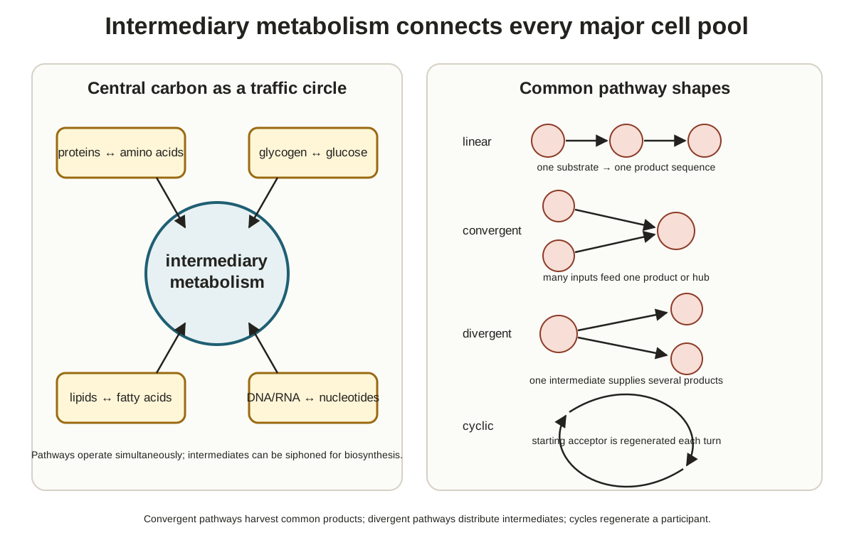
Small pools of energy-carrying compounds closely mesh catabolic breakdown with anabolic synthesis.
- Intermediary metabolism concerns small organic intermediates such as ATP, glucose, fatty acids, and amino acids rather than large proteins, DNA, or RNA. The lecturer placed them at about 1,000 molecular weight or less.
- Catabolic reactions break material down; anabolic reactions make cellular material.
- Nicotinamide adenine dinucleotide (
NAD) and its phosphorylated formNADPwere introduced alongside ATP,NADH, andFADH2as energy carriers or energy forms. - Cells do not store large quantities of these compounds. Their small pools force energy-producing breakdown and energy-using synthesis to remain closely connected.
- The battery and bank-account analogies emphasized that a cell does not maintain a huge reserve of ATP.
After a five-minute break, the lecturer restated the take-home point: cells are efficient, and when they build something, they must break something down.
Pathway Shapes Route Material Efficiently
Metabolic routes can converge, diverge, or cycle instead of running as many parallel pathways.
| Pathway shape | Routing described in the lecture | Example or qualification |
|---|---|---|
| Convergent catabolism | Many breakdown routes funnel toward one compound | Glucose through glycolysis, glycogen, and fatty acids were shown converging on acetyl-coenzyme A (acetyl-CoA) |
| Divergent anabolism | Routes branch like a tree while building material | Fatty-acid biosynthesis might be discussed at the end of the quarter, depending on class progress |
| Cyclic | A small pool of intermediates is reused | In the oversimplified example, a two-carbon acetyl-CoA input is balanced by two carbons leaving as CO2, so in theory the cycle could continue indefinitely |
- An acetyl group has two carbons. CoA was described as a carrier; the lecturer mentioned thioesters and immediately told students to set that added detail aside for now.
- Glycolysis begins with glucose and belongs to Midterm 1.
- Glycogen is a glucose polymer used for daily glucose storage.
- The Davis-to-San-Francisco analogy compared convergence with traffic funneling onto Interstate 80 rather than requiring parallel freeways.
- The cyclic example was the Krebs cycle. The lecturer revised “very complicated” to “not super complicated, but ... complicated.”
Exam emphasis: Know glycolysis for Midterm 1 like the back of one hand and the Krebs cycle for Midterm 2 like the other. The Krebs cycle is in mitochondria; glycolysis is in the cytosol.
Coupling Makes Glutamine Formation Proceed
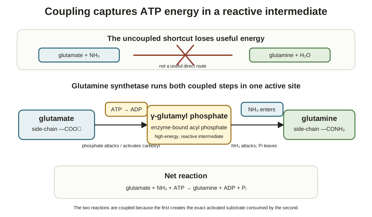
Glutamine synthetase physically couples endergonic glutamine formation to exergonic ATP breakdown at one active site.
- A student answer of aspartic acid was corrected to glutamic acid and then to the charged name glutamate, one-letter code
E. - The lecturer encouraged knowledge of one-letter amino-acid codes for broader biological and medical literacy but explicitly said this demand was not for this class.
- Glutamate (
E) reacts with ammonium to form glutamine (Q). - Free ammonium at high concentrations is toxic, although the lecturer said the reason is unknown. Nitrogen-containing cellular material can be broken down to ammonia, which becomes ammonium at cell pH.
- Some cells detoxify ammonium by converting it to harmless glutamine.
- Uncoupled glutamine formation is slow and endergonic. ATP breakdown to ADP and inorganic phosphate is exergonic, so coupling makes the overall process proceed in one direction.
Psubscripti, orPI, means inorganic phosphate and is acceptable notation on a test.
The two active-site steps
The millisecond-scale active-site process occurs in order:
- Activate glutamate. Glutamate and ATP bind specifically. The terminal phosphate of ATP is transferred only to glutamate's side-chain carboxyl group, not its other carboxyl group, forming an acyl-phosphate intermediate and ADP. Acyl phosphates are high-energy compounds; “high energy” here means a large negative
ΔGand energy release. - Add nitrogen. Ammonium enters, and an active-site group removes a proton to make ammonia. The nitrogen lone pair attacks the electron-poor carbonyl carbon, displacing phosphate. The ammonia supplies glutamine's
NH2, and inorganic phosphate is released.
What coupling does and does not require
- Coupling follows ordinary chemistry and can involve several molecules, often phosphate.
- Early discoveries of glycolysis and coupled reactions led biochemists to think everything in cells worked this way. The lecturer explicitly corrected that historical view as wrong and noted that proton-gradient mechanisms were discovered much later.
- Coupled reactions do not have to involve ATP; other compounds can participate. ATP is a classic example, and a similar case appears in glycolysis.
- In the lecturer's stated take-home, glutamate also serves ammonium detoxification, the ATP-derived phosphate creates the high-energy intermediate, and
E + ammonium -> Qwould not occur “ever” without the enzyme because the reactions are physically connected at its active site.
Glucose Must Enter Before Glycolysis
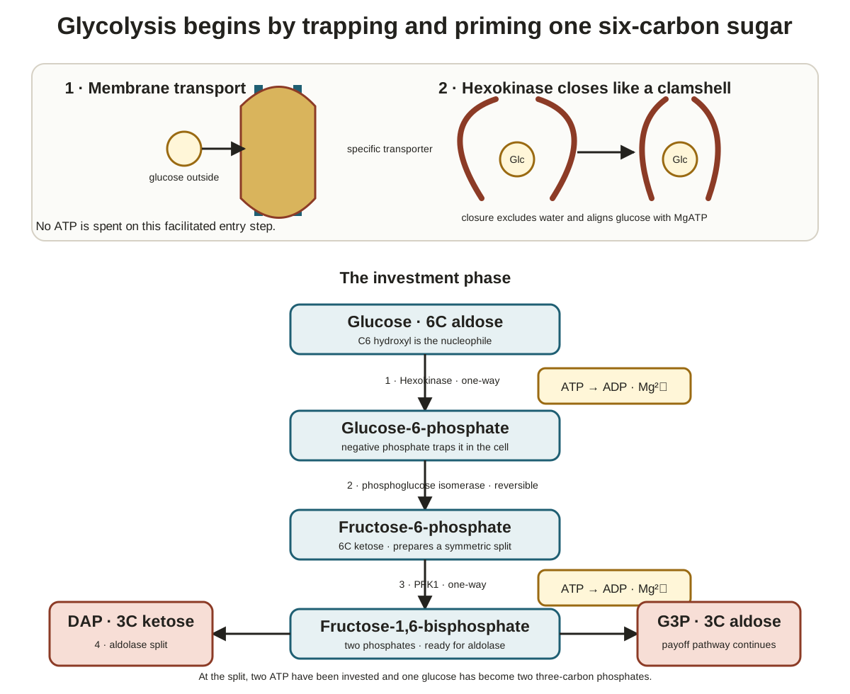
Glycolysis breaks a sugar in the cytosol, so polar glucose first requires a membrane protein to enter the cell.
- The course will focus on glucose rather than other sugars.
- The announced study lens includes the reaction sequence and its purpose, cellular efficiency, regulation of every pathway enzyme, and why glycolysis exists. Regulation was announced for later coverage rather than explained here.
- Alcoholic fermentation in some organisms was introduced as a slight change at the end of the pathway.
Beta-D-glucose conventions
- Glucose has six numbered carbons.
- Vertical shorthand lines are hydroxyl (
OH) groups, not methyl groups. - The carbon-2 hydroxyl must point down, and the carbon-3 hydroxyl must lie above the ring plane.
- The carbon-1 hydroxyl can lie above or below because the ring oxygen–carbon-1 bond is labile and the ring opens and closes in water. This is mutarotation.
- The beta form, with the carbon-1
OHabove the plane, was described as slightly more stable in solution. A horizontal bond at carbon 1 can represent either orientation.
Facilitated movement through the membrane
- Glucose is polar because it contains many oxygens. Oxygen, nitrogen, and sulfur were called polar atoms, and charge increases polarity.
- The plasma-membrane interior is nonpolar, so glucose needs protein help to cross.
- The lecturer described the glucose transporter as an integral membrane protein of about 45,000 molecular weight and said its X-ray structure and a step-by-step movie of glucose passage were not available.
- The transporter facilitates diffusion in both directions.
- Humans were described as having 14 forms, encoded by
GLUT1throughGLUT14, suited to different tissue and organ demands. The brain was offered as a high-demand example because it runs mostly on glucose.
Hexokinase Traps Glucose in the Cytosol
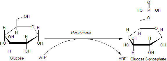
Hexokinase couples ATP use to phosphorylation of the carbon-6 hydroxyl, producing glucose 6-phosphate that cannot leave through the glucose transporter.
Glucose --[hexokinase; Mg cofactor; ATP consumed]--> glucose 6-phosphate
- Hexokinase is stereospecific but can act on sugars other than glucose.
- The lecturer described ATP going to ADP and inorganic phosphate and the phosphate being installed specifically on carbon 6.
- Circle-
Pshorthand is allowed on an exam. At physiological pH, the installed phosphate was depicted with a-2charge. - The first glycolysis step consumes one ATP even though one purpose of glycolysis is to produce ATP.
Four consequences of the carbon-6 phosphate
- Converting incoming glucose removes it and pulls more glucose inward.
- The glucose transporter recognizes glucose but not glucose 6-phosphate, so the product cannot escape.
- The phosphate provides an orienting “handle” for the next enzyme.
- Transferring some ATP energy raises glucose's energy slightly, although the lecturer emphasized that glucose already contains a great deal of energy.
Why magnesium is required
- Hexokinase specifically requires magnesium at the active site. Zinc, manganese, and other metal ions cannot substitute.
- The carbon-6
OHhas electron density and acts as a nucleophile toward ATP's partially positive phosphorus. The surrounding oxygens withdraw electrons, while ATP's negative-charge cloud hinders approach. Mg2+shields or dampens those negative charges so the nucleophile can approach. The lecturer said magnesium's exact position is still unknown.- Cytosolic water could also attack the phosphorus, but the glucose-
OHattack was stated to be 40,000 times faster.
Open and glucose-bound hexokinase
- X-ray evidence from yeast hexokinase showed a dimer, although only one identical subunit was displayed.
- A domain was defined as local tertiary, or local three-dimensional, structure. Each displayed subunit has two domains.
- Without glucose, the enzyme is open. Glucose binding triggers the domains to close like a clamshell and physically exclude water.
- Glucose alone does not permit catalysis because a second substrate is required, allowing the glucose-bound state to be crystallized and studied.

Monday Plan Before the Pathway Overview
Students were asked to learn two pathways, beginning with glycolysis, although the reactions and regulation would not all be covered that day.
- The lecturer planned to finish most of glycolysis on Monday.
The Two-ATP Investment Prepares a Three-Carbon Split
The opening phase phosphorylates the six-carbon sugar at carbon 6 and carbon 1, then aldolase splits between carbons 3 and 4 to produce two phosphorylated three-carbon groups.
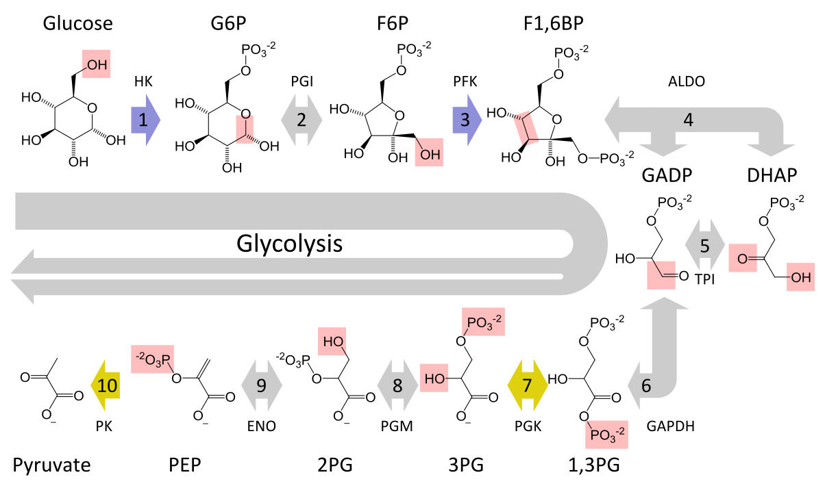

The carbon-handling plan
- The lecturer's overview ran from glucose through glycolysis to lactate, and Homework 1 requires learning the whole displayed sequence.
- In organisms such as yeast, alcoholic fermentation follows the pathway to pyruvate and then branches to ethanol.
- The cell first labels carbon 6 with phosphate, then carbon 1, and then cuts the six-carbon molecule between the two ends.
Return to the first ATP investment
- The carbon-6
OHacts as a nucleophile and attacks ATP. - Nucleoside triphosphates such as ATP and GTP contain substantial energy, but in the lecturer's framing they do not attack: they are attacked, and that releases energy.
Ring opening and phosphoglucose isomerase
- Mutarotation opens glucose 6-phosphate in water. The ring oxygen becomes an
OH, while carbon 1 becomes an aldehyde. - Aldehydes were described as good targets rather than good attackers.
- Phosphoglucose isomerase (
PGI) moves the bond to carbon 2, making carbon 1 an alcohol that can attack a later ATP. - PGI converts glucose 6-phosphate to fructose 6-phosphate in a totally reversible aldose/ketose isomerization. This contrasts with the preceding hexokinase step, which the lecturer called one-way.
The second ATP and aldolase split
PFK1, expanded in the lecture as “phosphofructokinase isomer 1,” consumes a second ATP and places phosphate at the other end. The lecturer explained the-1by noting thatPFK2was discovered later and will appear later in the course.- Two ATP molecules have now been consumed.
- Aldolase splits between carbons 3 and 4, producing dihydroxyacetone phosphate (
DAP) and “glyceroldehyde 3-phosphate” (G3P) in the transcript's wording.
Monday Continuation and Midterm Cutoff
The class ran out of time after the aldolase split, and the lecturer planned to finish most of glycolysis on Monday before drawing the first-midterm line.
- Students were told to learn the pathway over the four-day weekend.
- The lecturer promised to explain the logic of the rest of the pathway on Monday.
Study Review Questions
- How does living-cell microcalorimetry differ from combustion calorimetry, and which term in the Gibbs free-energy equation does it measure?
- Why must sodium hydroxide remain physically separate from the tissue, and why is it used in excess during Part B?
- What does each plateau in Parts A, B, and C test, and what would it mean if Part B equaled Part A?
- Why can the Part-B pressure decrease be assigned to oxygen consumption while pressure changes in Parts A and C cannot?
- How does dry-weight normalization make tissue samples comparable, and why is a vacuum used during drying?
- How do convergent, divergent, and cyclic pathways route metabolites differently?
- What ordered active-site steps allow glutamine synthetase to couple ATP breakdown to glutamine formation?
- How do carbon-6 phosphorylation, magnesium, and glucose-triggered clamshell closure contribute to the first glycolysis step?
- What sequence converts glucose 6-phosphate into the two products of the aldolase split, and where are the two ATP molecules consumed?