Lecture Overview
This lecture completed the Krebs cycle, covering the reactions from succinate through oxaloacetate, and then introduced coupled enzyme assays as a spectrophotometric technique for measuring enzyme activity. The three allosteric regulatory enzymes of the Krebs cycle were identified, and the cycle's roles as both an energy-producing and biosynthetic pathway were discussed. A detailed example of a coupled enzyme assay for acetate kinase was worked through, including the rationale for pH selection and dilution corrections. The final portion of the session was an open Q&A reviewing first midterm material.
Key Concepts and Definitions
- Succinate dehydrogenase: The only Krebs cycle enzyme attached to the inner mitochondrial membrane; transfers electrons to FAD; part of complex II of the electron transport chain.
- Fumarase: A catalytically perfect enzyme that adds water across the trans double bond of fumarate to produce L-malate.
- Catalytically perfect enzyme: An enzyme that cannot evolve to do its job better; catalyzes its reaction as fast as substrate contacts it.
- Anaplerotic reactions: "Filling up" reactions that replenish Krebs cycle intermediates removed for biosynthesis.
- Coupled enzyme assay: A spectrophotometric assay in which the reaction of interest is linked to one or more additional enzymatic reactions so that a measurable chromophore (such as NADH at 340 nm) is consumed or produced in proportion to the target reaction.
- Molar extinction coefficient: Numerically, the absorbance that would be measured for a one molar solution of a compound in a cuvette; larger values indicate more conjugated double bonds.
- Absorbance: Defined as the log base 10 of I0/I, where I0 is the incident light intensity and I is the transmitted light intensity.
- Ampholytes: Small molecules with different isoelectric points that generate the pH gradient in an IEF gel when voltage is applied.
Chronological Lecture Notes
Krebs cycle completion and roles
1. Course Logistics
- Short lecture day; no office hours, but the lecturer stayed briefly for questions.
- First midterm is tomorrow: last names A through N in the regular room, all others in 1215 TLC; 50 minutes; bring a calculator.
- Today's lecture finishes the material for the second midterm (September 2nd).
- Homework 2 will be posted Thursday, covering all second midterm material.
2. Krebs Cycle: Succinate Dehydrogenase and Complex II
- Picking up from the formation of succinate by succinyl-CoA synthetase (named for the nucleoside triphosphate involved).
- Succinate dehydrogenase transfers electrons to FAD (not NAD).
Exam emphasis: Succinate dehydrogenase is the only Krebs cycle enzyme attached to the inner mitochondrial membrane; all others float in the matrix.
- The enzyme's active site faces the matrix. Additional subunits connect it directly to the electron transport chain, embedded in the inner membrane.
- FADH2 does not diffuse away; electrons pass from the active site through the enzyme's subunits directly into the electron transport chain.
- Succinate dehydrogenase is part of complex II (Roman numeral II) of the electron transport chain.
- Product is fumarate (four-carbon compound) with a trans double bond: carboxyl groups are on opposite sides of the double bond, and protons are on opposite sides. The enzyme never makes the cis isomer.
3. Krebs Cycle: Fumarase and L-Malate
- Fumarase adds water across the trans double bond of fumarate.
- Always produces L-malate with the hydroxyl group on carbon 2 (drawn on the left).
- Fumarase is a catalytically perfect enzyme: it cannot evolve to perform its catalysis any better. The reason for this catalytic perfection is not currently understood.
4. Krebs Cycle: Malate Dehydrogenase and Thermodynamic Favorability
- Malate dehydrogenase uses NAD to oxidize L-malate to oxaloacetate (OAA), producing NADH and a proton.
- The conversion of L-malate to OAA is thermodynamically unfavorable (small positive or near-zero delta G).
- This does not prevent the pathway from functioning because the next reaction (citrate synthase, the first step of the Krebs cycle) is strongly one-directional with a large negative delta G, pulling the equilibrium forward.
- Not every reaction in a pathway needs to be thermodynamically favorable on its own; strongly favorable downstream reactions pull unfavorable ones forward.
- None of these reactions ever reach true equilibrium in the cell; they are always in an open, steady state, with products flowing into the next reaction.
- Once OAA is regenerated, a new acetyl-CoA molecule enters and the cycle repeats.
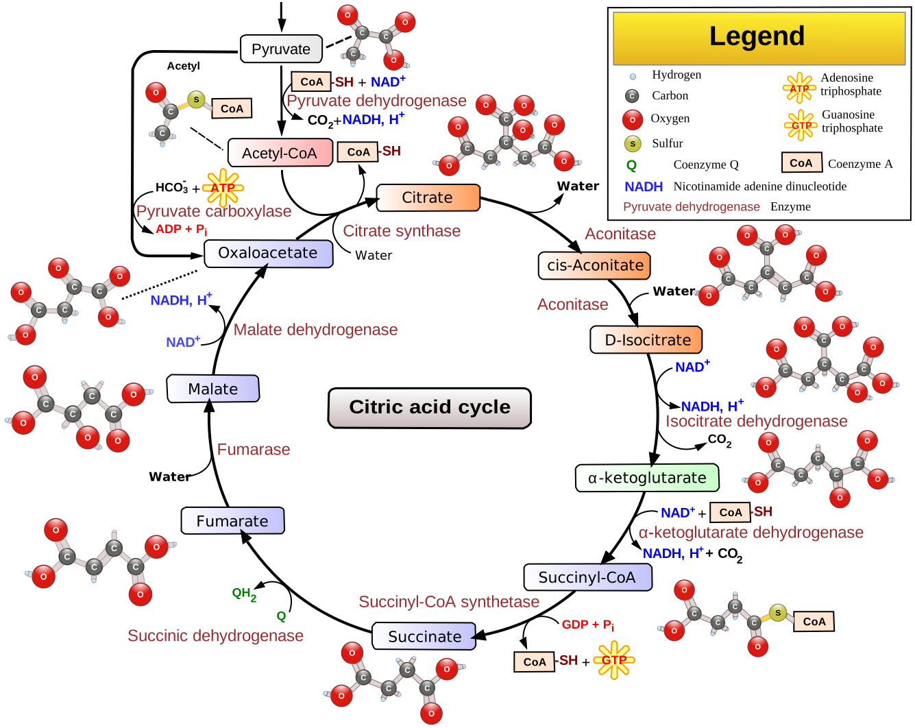

5. Allosteric Regulation of the Krebs Cycle
- Rule of thumb: reactions with large negative delta G values are usually controlled by allosteric enzymes.
- Three allosteric enzymes in the Krebs cycle:
- Citrate synthase — allosteric; velocity vs. substrate gives an S-shaped (sigmoidal) curve.
- Isocitrate dehydrogenase — one-way reaction driven by loss of CO2 (large negative delta G).
- Alpha-ketoglutarate dehydrogenase complex (enzymes E1, E2, E3) — driven by loss of CO2.
- The remaining Krebs cycle enzymes are Michaelis-Menten enzymes that give hyperbolic velocity vs. substrate plots.
6. Comparison of the Latter Krebs Cycle to Beta-Oxidation
- The last four steps of the Krebs cycle (succinate to oxaloacetate) parallel the four steps of beta-oxidation of fatty acids:
- A dehydrogenase uses FAD to introduce a trans double bond (like step 1 of beta-oxidation).
- Water is added across the double bond (like step 2 of beta-oxidation).
- A dehydrogenase uses NAD (like step 3 of beta-oxidation).
- A nucleophile attacks: in the Krebs cycle, the nucleophile is the carbon of the CH3 group (which becomes CH2 with a negative charge at the enzyme active site); in beta-oxidation, thiolase adds acetyl-CoA as the nucleophile.
7. Krebs Cycle as Biosynthetic Hub and Anaplerotic Reactions
- Krebs cycle intermediates (e.g., citrate, alpha-ketoglutarate) can be withdrawn to build other molecules the cell needs, similar to how glycolysis intermediates can be diverted.
- If carbons are removed for biosynthesis, anaplerotic ("filling up") reactions must replenish those carbons to keep the cycle running.
- In theory, the Krebs cycle runs indefinitely as long as acetyl-CoA enters (two carbons in) and two CO2 molecules leave per turn, but removing intermediates for biosynthesis requires anaplerotic input.
8. Roles of the Krebs Cycle
- Energy production for the electron transport chain:
- Three NADH molecules produced per turn go to complex I of the electron transport chain.
- FADH2 produced by succinate dehydrogenase goes directly into complex II.
- The electron transport chain is involved in generating the proton gradient in mitochondria.
- GTP production: GTP (guanosine triphosphate) is produced and is energetically equivalent to ATP. An enzyme can interconvert: GTP + ADP --> ATP + GDP. Some hormone receptors use GTP.
- Biosynthetic precursors: The cycle provides intermediates for building other molecules the cell needs.
Coupled enzyme assays
9. Coupled Enzyme Assays: Concept and Direct Assay Limitations
- Much biochemistry research has historically used radioisotopes, which work but are expensive to dispose of (long half-lives; must be shipped to specialized disposal facilities).
- Coupled enzyme assays use a spectrophotometer and are a cheaper alternative.
- Direct assay attempt for hexokinase:
Glucose + ATP --[Hexokinase]--> Glucose-6-phosphate + ADP- To use a spectrophotometer, something must absorb light, meaning it must have conjugated double bonds.
- Glucose has no conjugated double bonds and does not absorb.
- ATP absorbs at 260 nm (due to the adenine ring), so initial absorbance is high.
- Problem: ADP also absorbs at 260 nm with virtually the same intensity (same conjugated ring system, just missing a phosphate). As ATP is consumed and ADP is produced, absorbance does not change — the signal is canceled out.
- A flat (horizontal) absorbance line is uninformative: it could mean the enzyme is dead, a substrate was omitted, or the wavelength is wrong.
- Conclusion: directly assaying hexokinase at 260 nm does not work.
10. Coupled Enzyme Assay: Acetate Kinase Example
- Target reaction (bacteria only):
Acetate + ATP --[Acetate kinase]--> Acetyl phosphate + ADP- Same problem as hexokinase: ATP and ADP both absorb at 260 nm.
- Goal: Measure how much acetate kinase is present in a bacterial cell extract.
- Experimental setup: Grow cells (e.g., E. coli), break open in buffer, centrifuge to remove insoluble material (cell walls, membranes), and take a sample of the soluble extract.
- Coupling reactions:
- Add excess acetate and excess ATP (the substrates for the target enzyme).
- Add excess PEP (phosphoenolpyruvate) and excess pyruvate kinase: every ADP produced is immediately consumed.
ADP + PEP --[Pyruvate kinase]--> Pyruvate + ATP
- Add excess NADH and excess LDH (lactate dehydrogenase): every pyruvate produced is immediately consumed.
Pyruvate + NADH + H+ --[LDH]--> Lactate + NAD+
- NADH absorbs at 340 nm; nothing else in the assay absorbs at 340 nm.
- Three enzymes in the assay: Acetate kinase (the target being measured), pyruvate kinase (a tool), and LDH (a tool). The tool enzymes are added in excess.
- Stoichiometry: One-to-one throughout. Each ADP produced leads to consumption of one NADH molecule.
- Reading the assay:
- Monitor absorbance at 340 nm vs. time.
- Initial absorbance is very high because excess NADH is present.
- Absorbance decreases over time as NADH is consumed.
- Only the initial linear portion of the curve is used; the slope of this linear region equals the initial velocity (V0).
- Validation: If twice as much cell extract is added, the slope should be twice as steep. Plotting velocity vs. amount of acetate kinase added should give a linear relationship through the origin. A linear relationship confirms a valid coupled enzyme assay.
11. Coupled Enzyme Assays: pH Selection
- The three enzymes in the assay are unlikely to share the same optimal pH.
- Example: acetate kinase works best at pH 5, pyruvate kinase at pH 6, LDH at pH 7.
Exam emphasis: The buffer pH must be set to the optimum of the enzyme being measured (the target), not an average of all three. The tool enzymes can simply be added in greater amounts to compensate for suboptimal pH.
- The worst answer is to average the pH values (pH 6) — that reflects no understanding.
- The correct answer in this example is pH 5 (the optimum for acetate kinase, the target enzyme).
12. Coupled Enzyme Assays: Dilution Corrections
- In the homework problems, sometimes the goal is to assay a compound (e.g., fructose-6-phosphate) rather than an enzyme.
- General principle: try to extract as much information as possible from a single assay by pipetting in additional reagents.
- Practical pipetting: Adding a very small volume (e.g., 1 microliter) does not change the assay volume but introduces large pipetting errors. Adding a larger volume (e.g., 1 mL) is more accurate because a 1 microliter error on a 1 mL pipetted volume is negligible (1 part in 1,000), whereas a 1 microliter error on a 1 microliter pipetted volume is enormous.
- Dilution correction: If the original assay volume is 2 mL and 1 mL of reagent is added, the total volume becomes 3 mL. The original sample has been diluted by a factor of 2/3.
- To report the result in terms of the original undiluted sample, multiply by the inverse: 3/2.
- The correction factor returns the measurement to what it would have been without dilution (2/3 times 3/2 equals 1).
13. Second Midterm Material Complete
- The lecturer drew a line: all material for the second midterm (September 2nd) has now been covered.
- Electron transport chain will begin on Monday.
- Homework 2 will be posted Thursday.
Midterm review
14. First Midterm Q&A: Proton and Hydride Sources in Glycolysis
- The session shifted to answering student questions about first midterm material only.
- Student question: Where does the proton come from in the glyceraldehyde-3-phosphate dehydrogenase reaction?
- The proton comes from the phosphate group. At the relevant pH, the phosphate has the form with a P=O, two O- groups, and one OH; it is the OH that loses the proton.
- The hydride (H with both bonding electrons) that reduces NAD to NADH comes from the substrate carbon's C-H bond.
- Pyruvate to lactate example: NADH provides a hydride, which forms a new C-H bond on the carbon. The resulting double bond rearrangement produces an O- that grabs a proton from solvent to form the OH group.
- General principle: NADH always provides an H minus (hydride), and a proton from some compound or solvent is needed to maintain electrical neutrality.
15. First Midterm Q&A: Allosteric Enzymes in Glycolysis
- The one-way reactions in glycolysis (hexokinase, PFK1, pyruvate kinase) must be controlled and are allosteric enzymes.
- The other glycolysis enzymes are Michaelis-Menten enzymes — they respond less sensitively to regulation.
- Student question: Does ATP inhibit all three allosteric enzymes? As a rule of thumb, yes, but for the exam only PFK1 regulation was discussed in class. Asking about controls for hexokinase or pyruvate kinase specifically would not be fair.
- Many other compounds control allosteric enzymes, but those only make sense in the context of pathways not yet studied. There is extensive intercommunication between pathways.
16. First Midterm Q&A: Catalytically Perfect Enzymes in Glycolysis
- TPI (triosephosphate isomerase) is the only catalytically perfect enzyme in glycolysis.
- Glycolysis is one connected assembly line; the overall goal is to convert glucose all the way to pyruvate, which in eukaryotic cells goes into the mitochondria.
- Under exhaustion, lactic acid builds up and starts to slow things down; the system can only be pushed so far before energy sources run out.
17. First Midterm Q&A: Molar Extinction Coefficient
- The molar extinction coefficient is defined as the absorbance of a hypothetical one molar solution in a cuvette.
- For NADH, that value would be 6,220 — far too high for any spectrophotometer to measure, so in practice the sample is diluted to a measurable range, and absorbance scales proportionally.
- A larger molar extinction coefficient means the compound has more conjugated double bonds.
- Trade-off: more conjugated double bonds increase absorbance but also increase hydrophobicity, making the compound less water-soluble and harder to measure in aqueous buffers. Adding nonpolar solvents (ethanol, isopropanol) to dissolve such compounds risks denaturing enzymes.
18. First Midterm Q&A: Henderson-Hasselbalch Buffer Preparation
- Student question about preparing a Tris buffer at pH 7.3 (pKa = 8.21) from a stock solution at pH 9 using HCl.
- Cannot assume the stock at pH 9 is 100% base form because pH 9 is not two or more units above the pKa. Must use Henderson-Hasselbalch to find the base-to-acid ratio at pH 9.
- Then use Henderson-Hasselbalch again at pH 7.3 to find the new base-to-acid ratio. At 7.3 (well below the pKa), the acid form predominates.
- The difference in moles of base between the two states determines how much HCl to add.
- For a stock solution of H3PO4, the pH at the start does not need to be calculated — it is at a very low pH (nearly 100% H3PO4 form) and serves only as a starting point for titration upward. Calculating the exact starting pH wastes time.
19. First Midterm Q&A: LDH Isozymes and Gel Electrophoresis
- Bovine LDH is a tetramer of equal-sized subunits that come in two types: M (muscle) and H (heart).
- M subunits have a higher pI (more arginines and lysines, giving more potential positive charges).
- H subunits have a lower pI (fewer positives or more negatives).
- Five possible tetrameric combinations: M4, M3H, M2H2, MH3, H4.
- Different tissues produce different proportions of these forms: muscle (steak) is predominantly M4; heart is predominantly H4. Both tissues contain all five forms.
- IEF gel: Produces five bands, separated by pI. M4 bands at pI 8.2; H4 bands at pI 6. Does not provide size information.
- Native PAGE: Separates by size and charge-to-mass ratio. Subunits stay together. The positive electrode is at the bottom; tetramers with more H subunits (more negative charge) migrate fastest (H4 fastest). Size differences between the five tetramers are too small to resolve (within plus or minus 3,000 molecular weight). Running native gels at multiple resolving gel percentages (e.g., 6%, 8%, 10%) with protein standards allows calculation of native molecular weights.
- SDS-PAGE: SDS (sodium dodecyl sulfate, a strong detergent) completely denatures the protein. Mercaptoethanol reduces disulfide bonds. Only individual M and H subunits remain; since they have the same molecular weight, only one band appears.
- 2D gel (IEF then SDS): Five bands in the IEF dimension collapse to one band in the SDS dimension because all subunits have the same molecular weight.
- To confirm that all five IEF bands are LDH, each band must be excised and assayed for LDH activity (e.g., with pyruvate, NADH, and a proton).
- A protein with a lower pI may have more aspartates and glutamates, or fewer histidines, lysines, and arginines, or some combination.
20. First Midterm Q&A: Gel Polymerization and Ampholytes
- All gel types require polymerization: a free radical initiator (typically ammonium persulfate) starts the polymerization of acrylamide and bisacrylamide.
- Ammonium persulfate is a stable crystal; it begins generating free radicals when dissolved in water.
- A gel is like one giant cross-linked macromolecule with pores mostly filled with water. Without a gel matrix, electrophoresis in liquid would result in rapid diffusion that destroys band resolution.
- Too much acrylamide creates pores too small for proteins to enter. Typical gels are in the range of 6-8-10%; 30% gels are not used.
- IEF gels require ampholytes: small molecules with different isoelectric points that migrate to their pI positions when voltage is applied, establishing the pH gradient. Without ampholytes, there is no pH gradient.
Study Review Questions
- Why does FADH2 not diffuse away from succinate dehydrogenase, and how do its electrons reach the electron transport chain?
- Fumarase is described as catalytically perfect. What does this mean, and what stereochemical product does it always produce?
- If the conversion of L-malate to oxaloacetate is thermodynamically unfavorable, why does the Krebs cycle still proceed in the forward direction?
- Which three Krebs cycle enzymes are allosteric, and what thermodynamic feature do their reactions share?
- How do the last four steps of the Krebs cycle parallel the four steps of beta-oxidation?
- What are anaplerotic reactions, and why are they necessary for the Krebs cycle?
- Why does a direct spectrophotometric assay of hexokinase at 260 nm fail, and what property of the substrates and products causes this?
- In a coupled enzyme assay for acetate kinase, what is the role of pyruvate kinase and LDH, and why must they be added in excess?
- When multiple enzymes in a coupled assay have different pH optima, how should the buffer pH be chosen, and why?
- If a 2 mL assay receives a 1 mL addition of reagent, what dilution factor applies to the original sample, and what correction must be applied to the measured result?