Hormonal Regulation, Thiamine Pyrophosphate, and Pentose Phosphate Pathway
Date
2026-08-12
Source transcript
transcripts/en-BIS-103_ 2026-08-12 12_07.txt
Lecturer
Dr. Hilt
Subject Area
BIS-103 Biochemistry
Lecture Overview
This lecture covers three major areas. The first part examines how hormones (glucagon, epinephrine, and insulin) regulate metabolic pathways through signaling cascades, with emphasis on the role of fructose 2,6-bisphosphate in coordinating glycolysis and gluconeogenesis, G-protein-coupled receptor signaling, and the insulin receptor as a tyrosine kinase. The second part introduces thiamine pyrophosphate (TPP) as a coenzyme, explaining its three-part structure, its carbanion mechanism, and the disease beriberi. The third part covers the pentose phosphate pathway (oxidative and non-oxidative phases) and begins the transition into mitochondrial biochemistry with the pyruvate dehydrogenase (PDH) complex and coenzyme A.
Key Concepts and Definitions
Fructose 2,6-bisphosphate: A powerful allosteric effector made in small quantities; not part of glycolysis or gluconeogenesis. Stimulates PFK1 (glycolysis) and inhibits FBPase1 (gluconeogenesis).
PFK2/FBPase2: A bifunctional protein with two active sites on a single polypeptide chain. One site phosphorylates fructose 6-phosphate to fructose 2,6-bisphosphate; the other cleaves the phosphate off. An example of two fused genes.
PKA (Protein Kinase A): Also called cyclic AMP-dependent protein kinase A. Phosphorylates target proteins using ATP; activated by cyclic AMP binding to its regulatory subunits.
GPCR (G-protein-coupled receptor): A class of membrane receptors that use a G-protein trimer (alpha, beta, gamma subunits) to transmit signals. Approximately 350 unique GPCRs exist in humans.
Enzyme cascade response: A signaling amplification mechanism in which one hormone molecule can ultimately activate thousands of enzymes.
Cyclic AMP: A secondary messenger produced from ATP by adenylyl cyclase inside the cell.
Pro-glucagon / Pre-pro-insulin: Inactive precursor forms of hormones stored in cells; activated rapidly by enzymatic cleavage when needed.
Tyrosine kinase receptor: The class of receptor used by insulin; contains kinase domains that phosphorylate tyrosine residues. No G-protein or adenylyl cyclase involved.
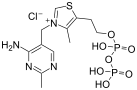
Thiamine pyrophosphate (TPP): A coenzyme with three essential parts (aminopyrimidine ring, thiazolium ring, pyrophosphate). Forms a carbanion that acts as a powerful nucleophile.
Ylid: A structure in which a carbanion (C-) is adjacent to a nitrogen; describes the reactive intermediate formed in TPP.
Beriberi: A serious disease caused by dietary lack of thiamine pyrophosphate, affecting multiple enzymes.
Pentose phosphate pathway: A pathway that converts glucose 6-phosphate into five-carbon sugars and NADPH. Has an oxidative phase and a non-oxidative phase.
NADPH: The phosphorylated, reduced form of NADP+; used as reducing power to maintain thiol (SH) groups and reduce other structures.
Glutathione: A tripeptide containing cysteine. When oxidized (disulfide form), it is inactive; glutathione reductase uses NADPH to regenerate active (reduced) glutathione.
Transketolase: An enzyme in the non-oxidative phase that moves two carbons; requires TPP.
Transaldolase: An enzyme in the non-oxidative phase that moves three carbons; uses a lysine-derived Schiff base, does not require TPP.
PDH complex (Pyruvate Dehydrogenase complex): A large multi-enzyme complex (~10 million molecular weight) in the mitochondrial matrix that converts pyruvate to acetyl-CoA.
Coenzyme A (CoA): A molecule with an SH group at its reactive end and an AMP handle. Forms high-energy thioesters. Discovered and synthesized by Professor Lippmann (Nobel Prize).
Thioester: A bond involving sulfur instead of oxygen; higher energy than an oxygen ester due to less resonance stabilization. Releases large negative delta G when broken.
Chronological Lecture Notes
Hormonal regulation of carbohydrate metabolism
1. Course Business and Exam Logistics
Answer key for homework one was posted before this lecture.
First midterm is next Wednesday; current lecture material will be on the second midterm.
Exam format: short answer, calculations, true-false. No Scantron.
Duration: 50 minutes. Students split between the lecture hall and a room on the first floor of the Teaching and Learning Center (TLC).
Pencil or pen acceptable; exams scanned via GradeScope. Write darkly if using pencil.
Bring a calculator.
2. Metabolic Control via Phosphorylation
Metabolism must be controlled; one mechanism is adding/removing phosphate groups on specific proteins.
Key examples discussed: phosphorylase and glycogen synthase.
3. Regulation of Glycolysis and Gluconeogenesis by Fructose 2,6-Bisphosphate
Gluconeogenesis and glycolysis run in opposite directions.
PFK1 and FBPase1 are both allosteric enzymes that are regulated.
PFK isomer 2 (PFK2) uses ATP to phosphorylate fructose 6-phosphate on carbon 2, producing fructose 2,6-bisphosphate. This is distinct from PFK1, which phosphorylates carbon 1 during glycolysis. Enzymes are stereospecific.
Fructose 2,6-bisphosphate has two phosphate groups, each with approximately minus 2 charge.
Fructose 2,6-bisphosphate is not part of glycolysis or gluconeogenesis; it is made in small quantities as an allosteric effector.
Allosteric enzymes produce an S-shaped curve when velocity is plotted against substrate concentration; fructose 2,6-bisphosphate can shift this curve left or right depending on the enzyme.
Fructose 2,6-bisphosphate binds PFK1 and stimulates it (accelerates glycolysis).
Fructose 2,6-bisphosphate binds FBPase1 and inhibits it (slows gluconeogenesis). Inhibition does not mean zero rate, just much slower.
One small organic molecule can simultaneously stimulate one pathway and inhibit the competing pathway.
To favor gluconeogenesis: break down fructose 2,6-bisphosphate. FBPase2 uses water to cleave the phosphate. Without fructose 2,6-bisphosphate, glycolysis loses its stimulation and gluconeogenesis loses its inhibition.
4. PFK2/FBPase2 as a Bifunctional Protein
PFK2 and FBPase2 activities reside on a single polypeptide chain with two active sites. This is a bifunctional protein resulting from evolutionary fusion of two genes.
Both enzyme activities are synthesized simultaneously, but only one active site is active at a time.
Phosphorylation of a particular serine (by PKA using ATP) switches which active site is active: one state favors PFK2 activity, the phosphorylated state favors FBPase2 activity.
The reverse reaction (dephosphorylation) is catalyzed by PP1 (phosphoprotein phosphatase 1), which uses water to remove the phosphate.
Exam emphasis: The lecturer stated students will never be asked to write out the full regulatory diagram on the second midterm or final; the focus is on the bigger picture and concepts.
5. Hormonal Control of PKA and PP1
PKA is stimulated by glucagon. Glucagon does not bind PKA directly; it binds a receptor in the cell membrane, triggering a series of events that stimulate PKA.
PP1 is stimulated by insulin. Insulin does not bind PP1 directly; it binds its own receptor.
Glucagon and epinephrine (adrenaline) inhibit PP1.
Hormones cause a cascade effect: one molecule of insulin, glucagon, or epinephrine can activate thousands of enzymes. This is not a one-to-one ratio; it is a massive amplification.
Edwin Krebs studied this phosphorylation and dephosphorylation system.
6. Glucagon: Structure and Function
Insulin and glucagon are both proteins, both made in the pancreas.
Fluorescence microscopy of mouse pancreas: insulin (green fluorescent probe) is made/stored in beta cells; glucagon (red) is made in alpha cells. Deep blue/purple marks nuclei.
Fluorescence microscopy uses UV excitation at 90 degrees to the viewer; fluorescent molecules glow at a longer wavelength against a dark background, providing greater detail than light microscopy.
Glucagon is 29 amino acids long; most of the backbone forms an alpha helix.
Synthesized as pro-glucagon (inactive precursor). An enzyme cleaves a piece off, changing its shape and activating it. Cells can store thousands of pro-glucagons and activate them rapidly.
Glucagon raises blood glucose concentration slowly (unlike epinephrine). Two mechanisms:
Promotes gluconeogenesis (making glucose from pyruvate).
7. GPCR Signaling Cascade
Epinephrine and glucagon bind different receptors, but both work through GPCRs.
The receptor is a protein integrated into the cell membrane. On the intracellular side sits a G-protein trimer with alpha, beta, and gamma subunits. The alpha subunit is designated G_s (stimulatory).
Approximately 350 unique GPCRs exist in humans, binding many different ligands.
Signaling sequence:
Hormone (e.g., epinephrine) binds receptor, causing a conformational change.
Alpha subunit releases GDP and picks up GTP (GTP is energetically similar to ATP but a different molecule).
Alpha subunit dissociates from beta and gamma subunits.
Alpha and gamma subunits are both membrane-anchored; alpha subunit slides along the membrane.
Alpha subunit binds and activates adenylyl cyclase, which converts ATP into cyclic AMP.
One hormone molecule can cause several trimers to dissociate, each alpha subunit activating a different adenylyl cyclase. Because these are enzymes, they rapidly convert many ATPs to cyclic AMP.
Cyclic AMP acts as a secondary messenger inside the cell.
Built-in shut-off: the alpha subunit has GTPase activity. GTP is hydrolyzed back to GDP plus phosphate, inactivating the alpha subunit. GTP acts as an on-off switch; while GTP is bound, the subunit is active, but the GTPase activity ensures it shuts off.
Professors Gilman and Rodbell received Nobel Prizes for discovering this system.
8. PKA Activation by Cyclic AMP
PKA consists of regulatory (R) and catalytic (C) subunits.
When cyclic AMP is absent, the R subunit sits in the active site (mouth) of the C subunit, sterically blocking catalytic activity.
One PKA molecule has four subunits: two R and two C.
Four cyclic AMP molecules bind to specific sites on the R subunits (two per R subunit), causing a conformational change that pulls the R subunits out of the C subunits.
Both C subunits become active. If only two cyclic AMPs are available, possibly only one C subunit is activated.
9. ACAP5 and Multi-Enzyme Complex Organization
ACAP stands for A-kinase-anchoring protein.
ACAP5 is anchored to the membrane via palmitoyl (palmitic acid) groups.
ACAP5 binds five different molecules/proteins: PIP3 (a phospholipid), the receptor protein, adenylyl cyclase, PKA, and a phosphatase. This is how it gets the "5" in its name.
This arrangement creates a multi-enzyme complex, bringing enzymes together for efficiency. When one enzyme converts substrate to product, the next enzyme immediately uses that product as its substrate.
PIP3 is a phospholipid with a glycerol backbone, fatty acids at positions 1 and 2, and an inositol group (an unusual sugar) linked via a phosphate ester. The inositol is phosphorylated; each phosphate carries a minus 2 charge.
10. Enzyme Cascade Amplification
One molecule of glucagon binding its receptor can ultimately lead to approximately 10,000 glucose molecules being released. This amplification across multiple enzymatic steps is called an enzyme cascade response.
In the liver, glucagon leads to release of glucose 1-phosphate. In muscle, epinephrine binds its own receptor for a similar cascade.
In this course, the three hormones of focus are glucagon, epinephrine (adrenaline), and insulin.
This ties back to the earlier discussion of phosphorylase A and phosphorylase B as different forms regulated by this cascade.
11. Insulin: Structure, Synthesis, and Function
X-ray structure: a dimer of two polypeptide chains (A chain and B chain), 51 amino acids total.
Contains multiple disulfide bonds. Rule of thumb: proteins with disulfide bonds are generally secreted outside the cell. Disulfide bonds provide extra covalent strength for proteins traveling through the blood.
Professor Sanger was the first person to sequence a protein (insulin) and received a Nobel Prize. He later became the first to sequence a nucleic acid (different method) and received a second Nobel Prize.
Synthesized in the pancreas as pre-pro-insulin (inactive precursor, more complex than initially thought). Cells build many copies of pre-pro-insulin; an enzyme can activate thousands within a minute or less by cleaving pieces, causing a conformational change to the active form.
Insulin reduces blood glucose. Sometimes called the "after-dinner hormone."
Two mechanisms:
Stimulates synthesis of glucose transport proteins, increasing glucose uptake from the blood into cells.
Stimulates glycogen synthesis to store excess glucose.
12. Insulin Receptor: Tyrosine Kinase
The insulin receptor does not use a G-protein or adenylyl cyclase. It is a receptor enzyme (tyrosine kinase receptor).
Structure: a tetramer with two alpha subunits (extracellular, detect insulin) and two beta subunits (span membrane, form intracellular kinase domains).
Each kinase domain contains three tyrosine residues involved in catalysis.
Without insulin: both kinase domains are inactive and do not phosphorylate anything.
Insulin binding causes a conformational shift in the beta subunits, analogous to the conformational shift in PKA when cyclic AMP binds. A portion of the peptide chain moves, activating the tyrosine residues.
The active kinase domains phosphorylate each other, then phosphorylate target enzymes.
Net result of insulin binding: stimulates biosynthesis of glucose transport proteins, stimulates glycogen synthesis, and affects many other processes.
Insulin affects over 90 genes in the human genome. Problems with insulin signaling underlie diabetes.
A protein domain is defined as local tertiary structure where part of a polypeptide chain folds into a compact unit.
TPP and the pentose phosphate pathway
13. Thiamine Pyrophosphate (TPP): Structure and Mechanism
TPP was previously mentioned as the coenzyme required by pyruvate decarboxylase in alcoholic fermentation (pyruvate to acetaldehyde + CO2). Multiple important enzymes use TPP.
Exam emphasis: Students will never need to draw the TPP structure but must be able to recognize it and know how it works.
TPP has three essential parts; removing any part renders the molecule nonfunctional:
Aminopyrimidine ring: Indirectly causes removal of a proton from a key carbon, enabling carbanion formation.
Thiazolium ring: Contains the reactive carbon (the "business end"). The adjacent nitrogen always carries a positive charge, which stabilizes the carbanion through a plus-minus interaction. This nitrogen acts as an electron sink.
Pyrophosphate group: Carries substantial negative charge at physiological pH. Positions the entire molecule precisely in the enzyme active site.
The carbanion formed on the thiazolium ring is a powerful nucleophile, better than any R group of the 20 amino acids.
Mechanism of carbanion formation: two hydrogens on the thiazolium ring sterically clash. A glutamic acid residue (in its acid form, COOH) in the enzyme donates a proton to a nitrogen on the aminopyrimidine ring. This triggers an electron rearrangement that, combined with the steric clash, causes the key proton to leave the carbon, forming the carbanion (C-). The resulting structure with C- adjacent to nitrogen is called an ylid.
The carbanion attacks a specific atom in pyruvate, releasing CO2 and ultimately producing acetaldehyde.
Reference figure · external sourceStructure of thiamine pyrophosphate (TPP), showing its three functional regions discussed in the lecture: the aminopyrimidine ring, the thiazolium ring containing the reactive carbon, and the pyrophosphate group. Source: Wikimedia Commons · Hbf878 · CC0 1.0 (Public Domain)
14. Enzymes Requiring TPP and Beriberi
Enzymes that require TPP in this course:
An enzyme in the pentose phosphate pathway (transketolase).
An enzyme in the pyruvate dehydrogenase (PDH) complex, located in the mitochondrial matrix.
An enzyme in the Krebs cycle, also in the mitochondrial matrix.
Beriberi is a serious disease, often fatal, caused by dietary lack of thiamine pyrophosphate. Originally discovered in cultures with rice-heavy diets; TPP is in the hull of the rice, which is removed during processing.
Modern fortification of foods (milk, bread) largely prevents beriberi. The main at-risk group today is alcoholics with poor diets, since alcohol contains no thiamine pyrophosphate.
Every disease is ultimately altered metabolism; removing this coenzyme affects multiple essential enzymes.
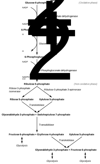
15. Pentose Phosphate Pathway: Overview and Oxidative Phase
Glucose 6-phosphate does not always enter glycolysis; it can enter the pentose phosphate pathway.
A pentose is a five-carbon molecule. Five-carbon sugars are needed for riboses and deoxyriboses in nucleic acids, ATP, NAD, and many other molecules. This pathway converts a six-carbon sugar into five-carbon products.
The pathway also produces NADPH, which provides reducing power. Cells need NADPH to maintain SH (thiol) groups. Disulfide bonds in the cytosol are generally undesirable because they inactivate molecules like coenzyme A. NADPH provides reducing power to break disulfide bonds back to functional thiols.
Disulfide bonds are appropriate for secreted proteins (e.g., insulin) and are generally made in the Golgi apparatus.
Glutathione is a tripeptide containing cysteine. When two glutathiones form a disulfide bond (oxidized form), they are inactive. Glutathione reductase uses NADPH to regenerate active (reduced) glutathiones.
Used to make riboses and deoxyriboses for DNA, RNA, and other molecules.
Carbons reshuffled through the non-oxidative phase to regenerate glucose 6-phosphate (if the cell needs more NADPH).
Six five-carbon molecules can be reshuffled into five six-carbon molecules. All reactions are reversible; the pathway can run in either direction depending on cellular need.
Transketolase: Moves two carbons from a donor to an acceptor. Requires TPP (carbanion mechanism).
Transaldolase: Moves three carbons from a donor to an acceptor. Does not require TPP. Uses a lysine residue in its active site to form a protonated Schiff base (N=C with positive charge) that carries the three-carbon unit.
In both enzymes, a nitrogen with a positive charge stabilizes a reactive carbanion.
Glyceraldehyde 3-phosphate and fructose 6-phosphate, both known from glycolysis, appear as intermediates in this reshuffling.
This pathway is called the oxidative pentose phosphate pathway. In photosynthesis, the Calvin cycle uses a reductive pentose phosphate pathway that consumes NADPH rather than producing it.
Reference figure · external sourceOverview of the pentose phosphate pathway, illustrating the oxidative phase (glucose 6-phosphate to ribulose 5-phosphate with NADPH production) and the non-oxidative phase (carbon reshuffling by transketolase and transaldolase). Source: Wikimedia Commons · Pink Bee · CC BY-SA 4.0
17. NADP+ Structure
NADP+ differs from NAD+ only by phosphorylation of an OH group on the ribose. That phosphate carries a minus 2 charge, allowing enzymes to distinguish between the two molecules.
Rule of thumb: the reduced form (NADPH) is used to reduce other things. A hydride (H-) is transferred to the top of the nicotinamide ring, just as with NAD+.
Mitochondria and the PDH complex
18. Transition to Mitochondria and Mitochondrial Structure
The pentose phosphate pathway and glycolysis occur in the cytosol. The course now moves into mitochondria.
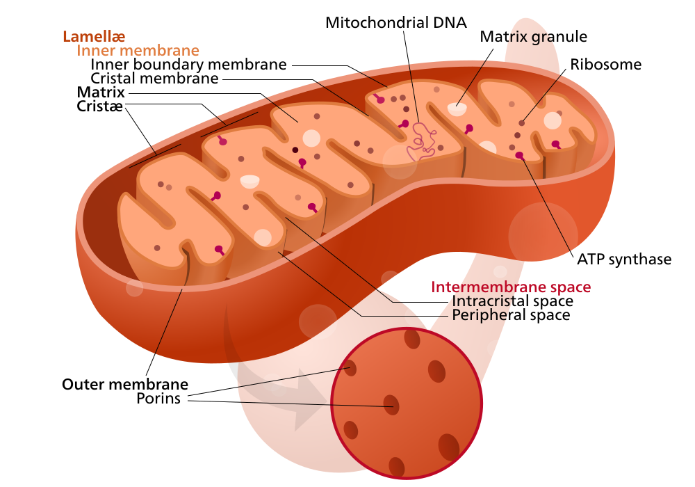
Mitochondrial structure:
Outer membrane (OM): Contains pores; molecules of approximately 5,000 molecular weight can freely cross between the cytosol and the intermembrane space. Pyruvate crosses easily.
Intermembrane space: Between the outer and inner membranes. Important later for pH gradients.
Inner membrane (IM): Convoluted. Nothing crosses unless a specific transport protein permits entry. Pyruvate has a dedicated transport protein in the inner membrane.
Matrix: The interior. Contains the PDH complex, Krebs cycle enzymes, beta-oxidation of fatty acids.
Reference figure · external sourceLabeled diagram of mitochondrial structure, showing the outer membrane, intermembrane space, convoluted inner membrane (cristae), and matrix discussed in the lecture. Source: Wikimedia Commons · Mariana Ruiz Villarreal (LadyofHats) · Public Domain
The electron transport chain (ETC) is embedded in the inner membrane. A typical mitochondrion has approximately 10,000 ETC complexes; in the heart, this can reach approximately 40,000.
In living cells, mitochondria do not look like textbook "potato" diagrams. They form a dynamic, interconnected network that constantly undergoes fusion (joining) and fission (dividing). This may ensure good mixing of mitochondrial contents.
Professor Nunnari on campus studied mitochondria for many years and was department chair before leaving to head an institute elsewhere.
Almost all the oxygen we breathe goes to the mitochondria.
19. PDH Complex: Reaction and Components
The PDH complex converts pyruvate into acetyl-CoA. It is a very large multi-enzyme complex (~10 million molecular weight) located in the mitochondrial matrix, containing interior tethers that facilitate the reaction.
Acetyl-CoA: CoA molecule with an SH group linked to a two-carbon acetyl unit via a thioester bond.
The complex contains many copies of three enzymes, designated E1, E2, and E3 in this course. Each requires a different coenzyme.
20. Coenzyme A, Thioesters, and FAD
Coenzyme A has an SH group at its reactive end ("business end") and a long chain leading to an AMP handle, similar in design to NAD+.
FAD (flavin adenine dinucleotide) also has an AMP handle; its business end is a tricyclic ring system.
Professor Lippmann earned a Nobel Prize for discovering coenzyme A and synthesizing it from scratch, confirming its structure.
Delta G-prime of acetyl-CoA hydrolysis is larger (more negative) than delta G-prime of ATP hydrolysis, meaning one molecule of acetyl-CoA contains more free energy than one ATP.
Thioesters are at a higher energy level than oxygen esters because there is less resonance stabilization between the carbonyl oxygen and sulfur compared to two oxygens. When a thioester breaks, it releases more energy.
Oxygen esters (e.g., fatty acid-glycerol linkages) are more stable and better suited for energy storage.
CoA must be in the SH (reduced) form to form a thioester.
Other CoA-linked molecules in this course: succinyl-CoA, fatty acid-CoAs. All carry thioesters and therefore contain substantial energy.
21. Closing Remarks
The lecturer stopped here, with plans to continue on Monday.
Students should study for the first midterm over the weekend.
Study Review Questions
How does fructose 2,6-bisphosphate coordinate glycolysis and gluconeogenesis, and what enzyme produces and degrades it?
Why can PFK2 and FBPase2 not be active at the same time, and what determines which activity is turned on?
Describe the signaling steps from glucagon binding its receptor to the production of cyclic AMP, including the role of the G-protein trimer.
How does the GTPase activity of the G-protein alpha subunit serve as an automatic shut-off mechanism?
What are the structural and mechanistic differences between the insulin receptor and GPCRs used by glucagon and epinephrine?
What are the three structural parts of thiamine pyrophosphate, and what role does each play in generating the reactive carbanion?
What are the two main products of the pentose phosphate pathway, and under what cellular conditions would the non-oxidative phase regenerate glucose 6-phosphate?
Compare transketolase and transaldolase in terms of the number of carbons transferred and cofactor requirements.
What are the substrates, products, and coenzymes of the pyruvate dehydrogenase complex reaction?
Why do thioesters contain more free energy than oxygen esters, and what is the biological consequence of this difference?