Lecture Overview
This lecture began with interactive exam review exercises covering phosphate buffers, IU calculations for LDH, and allosteric enzymes in glycolysis. The main new content covered how glycogen branches are formed and subsequently broken down by phosphorylase and the bifunctional debranching enzyme, with debranching as the rate-limiting step. The lecture then introduced gluconeogenesis as a high-energy pathway that is not a simple reversal of glycolysis, highlighting the roles of mitochondrial CO2, the biotin tether on pyruvate carboxylase, and the malate shuttle for transporting reducing equivalents. The session concluded with an introduction to enzyme regulation by phosphorylation and dephosphorylation, illustrated through glycogen synthase and phosphorylase.
Key Concepts and Definitions
- Branching enzyme: Moves groups of glucose residues to create alpha-1,6 linkages, generating new non-reducing ends on glycogen.
- Limit dextrin: The structure remaining after phosphorylase can no longer cleave glucose residues near branch points.
- Debranching enzyme: A bifunctional protein (single polypeptide, two active sites) that removes glycogen branches via transferase and glucosidase activities.
- Bifunctional protein: A single polypeptide chain with two different enzyme active sites, one N-terminus and one C-terminus, resulting from two genes that fused together in evolution.
- Phosphorylase: Enzyme that removes glucose residues from the non-reducing end of glycogen using inorganic phosphate, producing alpha-glucose-1-phosphate.
- Gluconeogenesis: The synthesis of glucose from scratch, starting from pyruvate; occurs mainly in the cytosol, begins in mitochondria, ends in the ER.
- Oxaloacetate (OAA): Four-carbon molecule formed from pyruvate + CO2 by pyruvate carboxylase; also a member of the Krebs cycle.
- Tether: A covalent linkage (e.g., biotin + lysine side chain) that carries a substrate between active sites within an enzyme, maintaining high local molarity.
- Biotin: A coenzyme (vitamin) that carries CO2; linked to the epsilon amino group of a lysine residue on pyruvate carboxylase.
- Isozymes: Different forms of the same enzyme with different primary structures (amino acid sequences), folded differently and evolved for different compartments.
- Shuttle system: A mechanism using a molecule (e.g., malate) to carry electrons indirectly out of an organelle when NADH itself cannot cross.
- Fructose 2,6-bisphosphate: An isomer of fructose 1,6-bisphosphate; a powerful allosteric regulator not part of glycolysis or gluconeogenesis, used as a controlling molecule.
- GSK3 (glycogen synthase kinase 3): Enzyme that phosphorylates serines on glycogen synthase, reducing its activity.
- PP1 (phosphoprotein phosphatase 1): Enzyme that uses water to remove phosphates from proteins, restoring their unphosphorylated state.
- Insulin: Protein hormone that binds to a membrane receptor; net effect is to inhibit GSK3 and stimulate PP1, removing glucose from the blood.
- Glucagon: Protein hormone that binds to a membrane receptor; inhibits PP1, putting glucose into the blood slowly.
- Epinephrine (adrenaline): Fight-or-flight hormone; also inhibits PP1, putting glucose into the blood fast.
Chronological Lecture Notes
Glycogen breakdown
1. Opening and exam review exercises
- Midterm is a week from tomorrow (Wednesday). Some students will take the test in the lecture hall; others in the Teaching and Learning Complex (TLC) due to alternate seating.
- Interactive exercise: how to make a phosphate buffer at pH 7 starting with K3PO4.
- Phosphoric acid has a titration curve; K3PO4 starts at the very top.
- To come down to pH 7, add HCl.
- First step: add one equivalent of HCl to transform all PO4^3- to HPO4^2-.
- Next step: use the Henderson-Hasselbalch equation with the pKa of 7.21 (would be given on the exam).
- Solve for A (acid), because after the first step you have all B (base); you need to move some B over to A.
- Then add the total amount of acid.
- Interactive exercise: how to calculate IUs (international units) of LDH activity from a cell extract.
- Run the LDH assay (e.g., using pyruvate and NADH).
- Measure A340 as a function of time to get change in absorbance per unit time.
- Convert to change per minute (IUs require per-minute units).
- Use Beer's Law with the extinction coefficient of 6,220.
- Calculate molar change of NADH per minute.
- Multiply by the volume of the assay in liters (e.g., 2 mL = 0.002 L) to cancel liters from the molar concentration.
- Convert moles to micromoles to obtain IUs.
- Interactive exercise: identify the allosteric enzymes in glycolysis and write balanced chemical reactions for them.
Exam emphasis: None of these three types of questions are multiple choice. Bring a calculator. Students should be prepared to write out the entire balanced pathway of glycolysis with structures, enzyme names, and which reactions are unidirectional or reversible.
- Answer key for homework will be posted tomorrow; students should attempt problems first.
2. Glycogen branching
- Material for the second midterm begins here.
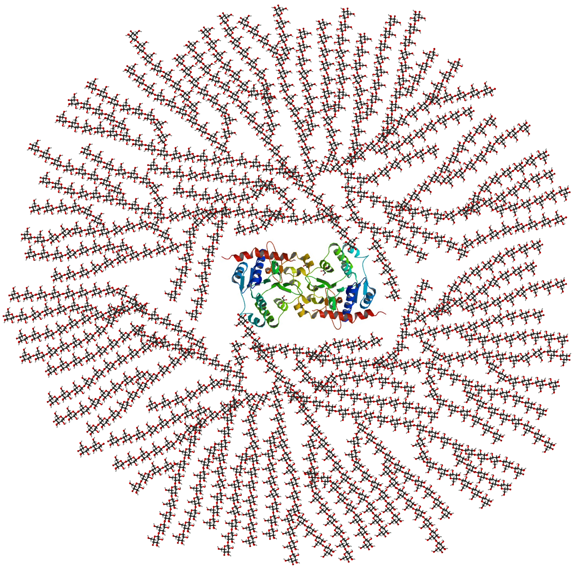
- Glycogen is a storage molecule in animals that stores glucose with lots of branches and non-reducing ends.
- All internal linkages are alpha-1,4; branches are alpha-1,6 linkages.
- The branching enzyme moves a group of glucose residues from one strand and reattaches them via an alpha-1,6 linkage, creating two non-reducing ends where there was one.
- Glycogenin is the protein dimer that serves as the anchor at the core.
- Glycogen grows like a tree with successive tiers of branching: each tier branches off previous branches.
- The number of non-reducing ends grows exponentially with each tier.
- All biochemistry (addition and removal of glucose residues) occurs at the non-reducing ends.
- A mature glycogen molecule has 12 tiers and approximately 50,000 glucose residues.
- Molecular weight is approximately 9 million (about 180 grams per glucose residue times 50,000).
- Glycogen cannot be seen without staining because glucose molecules lack conjugated double bonds.

3. Reasons for storing glucose as a polymer
- Reason 1: Polymerization protects glucose from oxidation. Individual glucose residues in the polymer cannot open and close, so they remain as functional glucose rather than oxidized derivatives.
- Reason 2: Reduces osmotic pressure. Osmotic pressure depends on the number of particles. One glycogen molecule replaces approximately 50,000 free glucose molecules, reducing osmotic pressure by about 50,000-fold.
- Water spontaneously moves from high concentration to low concentration across membranes.
- Plants solve the resulting pressure with cell walls; animals must solve it differently (e.g., pumping water out costs energy, or storing glucose as a polymer).
4. Glycogen breakdown by phosphorylase
- Phosphorylase attacks the non-reducing end, breaking the glycosidic bond and adding inorganic phosphate to produce alpha-glucose-1-phosphate (phosphate at carbon 1 in the alpha position).
- Using inorganic phosphate instead of water avoids the need to spend an ATP later to phosphorylate free glucose (as hexokinase does in glycolysis).
- Inorganic phosphate is very prevalent in cells.
- Once a phosphate is connected to the hydroxyl group at carbon 1, the ring cannot open and close.
5. Glucose-1-phosphate to glucose-6-phosphate
A mutase converts glucose-1-phosphate to glucose-6-phosphate by moving the phosphate from position 1 to position 6.
- Analogous to the 3-PGA to 2-PGA conversion seen in glycolysis.
Glucose-6-phosphate can enter glycolysis or the pentose phosphate pathway.
Student question about glucose sources: cells can take in free glucose molecules, but often we eat polymers that are broken down by digestive enzymes. There are also redundant pathways for making glucose (e.g., converting mannose).
6. Phosphorylase nibbling and the limit dextrin problem
- Phosphorylase sequentially removes glucose residues from non-reducing ends but cannot handle residues near the alpha-1,6 branch point because they do not fit into its active site.
- The remaining structure after phosphorylase stalls is called the limit dextrin.
7. The debranching enzyme
- The debranching enzyme rescues the limit dextrin situation. It is a bifunctional protein: a single polypeptide chain with one N-terminus and one C-terminus, containing two different active sites.
- Contrast with LDH, which has four subunits each with the same active site.
- The debranching enzyme arose from two genes that fused in evolution; when the protein folds, both activities are ready.
- Transferase activity: moves three glucose residues from the branch stub to another glycogen strand (alpha-1,4 linkages).
- Glucosidase activity: breaks the single remaining alpha-1,6 linkage, releasing the last glucose residue of the branch.
- Removing branches is the rate-limiting step of glycogen breakdown; phosphorylase nibbling is very fast by comparison.
- The balance of branching: more branches means more non-reducing ends (faster glucose release and addition), but also more limit dextrins requiring debranching. This balance is presumably why branching occurs every eight residues rather than every three.
8. Glycogen in cells and storage capacity
- Electron microscopy of rat liver shows glycogen granules as small dark spots (when stained). Groups of glycogen molecules linked together form rosettes.
- A starved rat would have no visible rosettes.
- Humans can store glycogen for only about one day; glycogen levels constantly fluctuate depending on eating and exercise.
- Student question about regulation: everything is regulated; loss of regulation is associated with serious disease.
Gluconeogenesis
9. Introduction to gluconeogenesis
- Gluconeogenesis: building glucose from scratch, needed when glycogen is depleted (starvation, overnight).
- The brain still needs glucose even when glycogen is gone.
- Occurs in some cells in animals, not all. The reactions occur mainly in the cytosol, start in the mitochondria, and end in the ER.
- Requires a large energy investment because glucose contains a great deal of energy.
Exam emphasis: The lecturer will never ask students to write out the pathway of gluconeogenesis, but they must understand the concepts.

10. Gluconeogenesis vs. glycolysis
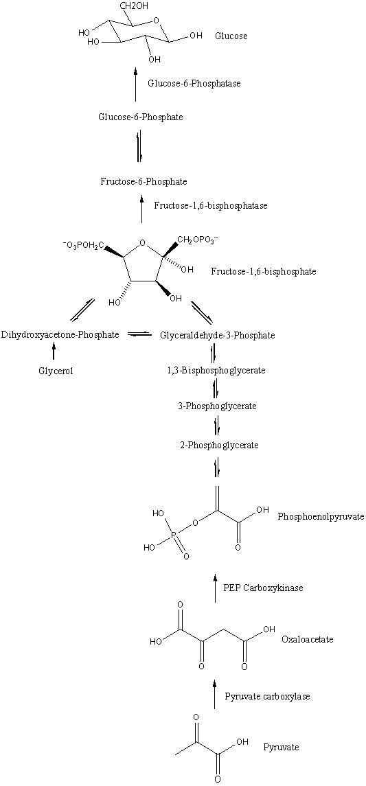
- Many of the enzymes in the middle of glycolysis catalyze reversible reactions; these same enzymes work in the reverse direction for gluconeogenesis. The cell does not need to make new enzymes for these steps.
- The three allosteric enzymes in glycolysis catalyze very large negative delta G reactions. These steps cannot simply be reversed; gluconeogenesis must use different enzymes to bypass each one.
- The step from PEP to pyruvate in glycolysis releases so much energy that it takes two steps to reverse it in gluconeogenesis.
- Waterfall/kayaking analogy: irreversible glycolytic steps are like waterfalls. You cannot kayak up; you must portage around them with different enzymes and different chemistry.
11. Gluconeogenesis: pyruvate to PEP
- Glucose is six carbons, so two pyruvates (three carbons each) are needed, each requiring its own energy investment.
Step 1 (in the mitochondrion): Pyruvate --[Pyruvate carboxylase; ATP, CO2]--> Oxaloacetate (OAA)- Mitochondria produce abundant CO2 from other reactions; CO2 is small, nonpolar, diffuses fast, hard to capture in the cytosol.
- OAA is a four-carbon molecule, also a member of the Krebs cycle.
Step 2 (in the cytosol): OAA --[PEP carboxykinase; GTP]--> PEP + CO2- GTP is energetically equivalent to ATP.
- The CO2 added in step 1 is released in step 2 to help drive the reaction thermodynamically (product escape pulls the reaction forward).
- The combined energy input (ATPs and GTPs) to make two PEPs is substantial.
12. Remaining gluconeogenesis steps and compartmentalization
- PEP carboxykinase and most remaining reactions occur in the cytosol.
- New enzymes specific to gluconeogenesis: PEP carboxykinase, fructose 1,6-bisphosphatase isomer 1 (FBPase-1).
- Most other reactions use the same reversible glycolytic enzymes.
Last step: Glucose-6-phosphate --[Glucose-6-phosphatase]--> Glucose- Glucose-6-phosphatase is located inside the ER.
- This makes sense because cells performing gluconeogenesis (mainly liver) send glucose elsewhere in the body (e.g., to the brain), and the ER is part of the cell's packaging and export system.
13. Allosteric control in gluconeogenesis and exceptions
- Pyruvate carboxylase: allosteric enzyme (also has a tether).
- Fructose 1,6-bisphosphatase isomer 1 (FBPase-1): allosteric enzyme.
- PEP carboxykinase: catalyzes a one-way reaction but is not allosterically controlled; it is regulated at the DNA level (gene expression controlling how many copies are made). This is an exception to the allosteric rule of thumb.
- Glucose-6-phosphatase: catalyzes a one-way reaction but is in a different compartment (ER), another exception.
- Most exceptions to the rule of thumb for allosteric control occur in gluconeogenesis.
- Competing pathways (glycolysis down, gluconeogenesis up) cannot both run at maximum velocity simultaneously.
14. The tether concept: pyruvate carboxylase and biotin

- Pyruvate carboxylase is the first example of a tether in the course.
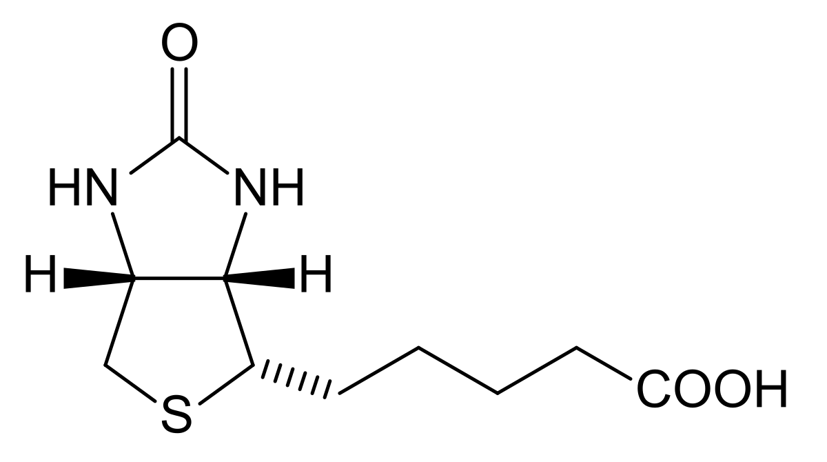
- Biotin is a coenzyme (small organic molecule needed for enzyme activity) and a vitamin.
- Almost all coenzymes are vitamins.
- Biotin is linked to the epsilon amino group of a specific lysine residue on pyruvate carboxylase by another enzyme.
- The tether consists of biotin plus the lysine side chain together, forming a rope-like structure.
- CO2 is covalently attached to a nitrogen on biotin. The tether carries the CO2 from one active site region to another within the enzyme.
- The tether keeps the CO2 at a very high local molarity. Without the tether, the cell would need much more biotin in free solution.
- Tetherball analogy: the rope can only swing a fixed distance from the pole.
- CO2 molecules are tiny and diffuse very fast; covalently attaching CO2 to the tether prevents escape.
- At the second active site, pyruvate attacks the CO2 to form oxaloacetate.
- More tethers will appear later in the course (PDH complex, Krebs cycle).
15. Mitochondrial compartmentalization and the malate shuttle
- Pyruvate is small enough to pass through pores in the outer mitochondrial membrane (cutoff approximately 5,000 molecular weight) into the intermembrane space, but requires a transport protein to cross the inner membrane into the matrix.
- In the cytosol, the G3PDH (glyceraldehyde-3-phosphate dehydrogenase) step of glycolysis is reversible and uses NAD to make NADH. Going backward in gluconeogenesis, this step needs NADH in the cytosol.
- Mitochondria have their own separate pools of NAD/NADH and do not share them with the cytosol.
- Shuttle system: instead of sending NADH out, the mitochondria transfer electrons onto oxaloacetate to make malate. Malate exits the mitochondria and is reoxidized in the cytosol, regenerating NADH there.
OAA + NADH --[Malate dehydrogenase (mitochondrial)]--> MalateMalate --[Malate dehydrogenase (cytosolic)]--> OAA + NADH
- The two forms of malate dehydrogenase (mitochondrial and cytosolic) are isozymes: different primary structures (amino acid sequences), different folding, each evolved for the conditions of its compartment.
16. Gluconeogenesis energy summary and conclusions
- Overall input for one glucose: 2 pyruvates, 4 ATP, 2 GTP, 2 NADH, protons, 4 waters.
- NADH, GTP, and ATP are all forms of energy. The total energy cost is enormous, but there is no alternative.
- The energy for gluconeogenesis comes from breaking down other molecules, primarily fatty acids. In a worst-case scenario, it can come from breaking down proteins.
- Gluconeogenesis is not the complete reversal of glycolysis; irreversible steps must be bypassed with different enzymes.
Regulation by phosphorylation
17. Introduction to regulation: levels of control
- Enzymes can be Michaelis-Menten or allosteric, and both types have inhibitors.
- Additional levels of control beyond allosteric regulation:
- Phosphorylation and dephosphorylation of enzymes
- Fructose 2,6-bisphosphate (a powerful allosteric regulator, isomer of fructose 1,6-bisphosphate, not part of glycolysis or gluconeogenesis)
- Hormones (chemical messengers)
- Edwin Krebs, a professor at UC Davis in the biological chemistry department (Tupper Hall), received the Nobel Prize for discovering enzyme regulation by phosphorylation and dephosphorylation. This is a different person from the Krebs who discovered the Krebs cycle.
18. Phosphorylation: which residues and how
- Three amino acids can be phosphorylated: serine (S), threonine (T), and tyrosine (Y). All three share an OH group in their side chains.
- Which specific residue is phosphorylated depends on the enzyme; even if an enzyme has many serines, typically only one gets phosphorylated.
- At physiological pH, a phosphate group carries a charge of about minus two, compared to no charge on the unmodified hydroxyl. This significantly affects the enzyme.
- A kinase uses a nucleoside triphosphate to add a phosphate. A phosphatase uses water to remove a phosphate.
19. Glycogen synthase regulation by phosphorylation
- Glycogen synthase: dimer with multiple serines on each subunit.
- Active form: unphosphorylated.
- GSK3 (glycogen synthase kinase 3) uses up to 6 ATPs to phosphorylate up to 6 serines, making glycogen synthase less active.
- Degree of inactivation is on a spectrum: more phosphates mean less activity. A population of glycogen synthase molecules may have varying numbers of phosphates.
- PP1 (phosphoprotein phosphatase 1) uses water to remove phosphates, converting the less active form back to the active form.
- Insulin (protein hormone): binds to a membrane receptor (not directly to GSK3 or PP1). Net effects: inhibits GSK3 and stimulates PP1.
- Glucagon (protein hormone): binds to a receptor. Net effect: inhibits PP1.
- Epinephrine (adrenaline): also inhibits PP1.
- Hormones bind to receptors and trigger intracellular chain reactions; they do not bind directly to these enzymes.
- Big-picture hormone effects on blood glucose:
- Insulin removes glucose from the blood.
- Glucagon puts glucose into the blood slowly.
- Epinephrine puts glucose into the blood fast (fight-or-flight).
20. Phosphorylase regulation by phosphorylation
- Phosphorylase: dimer with one serine per subunit.
- Active form: phosphorylated (called phosphorylase A). This is the opposite of glycogen synthase.
- Inactive form: dephosphorylated (called phosphorylase B).
- PP1 removes phosphates (phosphorylase A to B), inactivating the enzyme.
- Phosphorylase B kinase uses ATP to phosphorylate serine (phosphorylase B to A), activating the enzyme.
- Glucagon and epinephrine stimulate phosphorylase B kinase.
Exam emphasis: Phosphorylation does not always mean inactivation. For glycogen synthase, phosphorylation inactivates. For phosphorylase, phosphorylation activates. It depends on the enzyme.
Study Review Questions
- What are the two reasons cells store glucose as a polymer (glycogen) rather than as free glucose molecules?
- How does phosphorylase differ from a hydrolytic enzyme in the way it cleaves glucose residues from glycogen, and what is the energetic advantage of this mechanism?
- Describe the two activities of the debranching enzyme and explain why it is called a bifunctional protein.
- Why is removing branches the rate-limiting step of glycogen breakdown, and how does this relate to the frequency of branching in glycogen?
- Why can gluconeogenesis not simply be the reverse of glycolysis? What must the cell do at each irreversible glycolytic step?
- Why does the first step of gluconeogenesis (pyruvate carboxylase) occur in the mitochondria rather than the cytosol?
- Explain the tether concept using pyruvate carboxylase and biotin as an example. What advantage does the tether provide?
- How does the malate shuttle solve the problem of supplying NADH to the cytosol for gluconeogenesis when mitochondria do not export NADH directly?
- Compare the effects of phosphorylation on glycogen synthase versus phosphorylase. Why is it important not to assume phosphorylation always activates or always inactivates an enzyme?
- What are the respective effects of insulin, glucagon, and epinephrine on blood glucose levels, and how do these hormones exert their effects on intracellular enzymes?