DNA Microarrays and Two-Dimensional Gel Electrophoresis
Date
August 4, 2026
Source transcript
transcripts/en-BIS-103_ 2026-08-04 12_07.txt
Lecturer
Dr. Hilt
Subject Area
Biochemistry
Lecture Overview
The lecture completed prerequisite reviews of affinity chromatography, analytical protein gels, fatty acids, and two major lipid classes before applying those ideas to methods that survey cellular metabolism. DNA microarrays were developed as a two-sample workflow that isolates messenger RNA, converts it to differently colored complementary DNA, and measures competition at gene-specific spots. Two-dimensional isoelectric focusing/SDS-PAGE was then developed as a complementary method that separates a proteome first by isoelectric point and then by subunit size. Microcalorimetry appeared in the course-map scope but was not taught; it was explicitly deferred to the next class.
Course Expectations and Affinity-Column Review
Homework 1 defines the assignment scope for the first midterm, after which the lecture returned to affinity chromatography as a prerequisite for the day’s metabolic methods.
Homework 1 had been posted and was to be discussed at the first break. It is the only assignment for the first midterm, covers many topics, and was posted early so its scope would not surprise students just before the exam.
Accommodated Exam Services had confirmed arrangements. Its stated difference was that an accommodated exam must be taken between 8 a.m. and 5 p.m. on the same day as the class exam; the lecturer guessed that the cutoff reflected staffing limits.
How an affinity column selects its target
The microscopic beads are polar and porous, with pores large enough for all biomolecules to enter and leave.
The lecturer emphasized a correction: the bead is not a molecular sieve and does not separate molecules by size.
An organic arm is covalently attached to the bead, and ligand L is covalently attached to the arm, so the ligand cannot come off.
A six-carbon arm was recommended.
One- or two-carbon arms are too short to lift the ligand adequately.
Arms of 12 carbons or longer could make the bead behave as though it were hydrophobically coated.
The ligand is usually a small organic molecule and is often a substrate. It could instead be a product, inhibitor, coenzyme, or metal-ion cofactor, depending on what the active site recognizes.
Hexokinase, identified here as the first enzyme in glycolysis, uses glucose and ATP. Supplying both on a column would allow catalysis, so only one substrate is supplied to permit binding without reaction.
Hundreds of ligands can occur inside and outside each bead and are free to rotate. In a buffer at an appropriate pH, the target enzyme recognizes a ligand at its active site through one or more weak interactions:
ionic bonds
hydrogen bonds
van der Waals interactions
the hydrophobic effect
Each individual interaction is dynamic and weak, but repeated binding opportunities retain the enzyme noncovalently while material that does not recognize the ligand flows through.
This isolation logic matters because cellular components must be separated to study metabolism, and one or more affinity columns are used in the DNA-microarray procedure taught later in the lecture.
Analytical Gels Preserve or Remove Different Protein Features
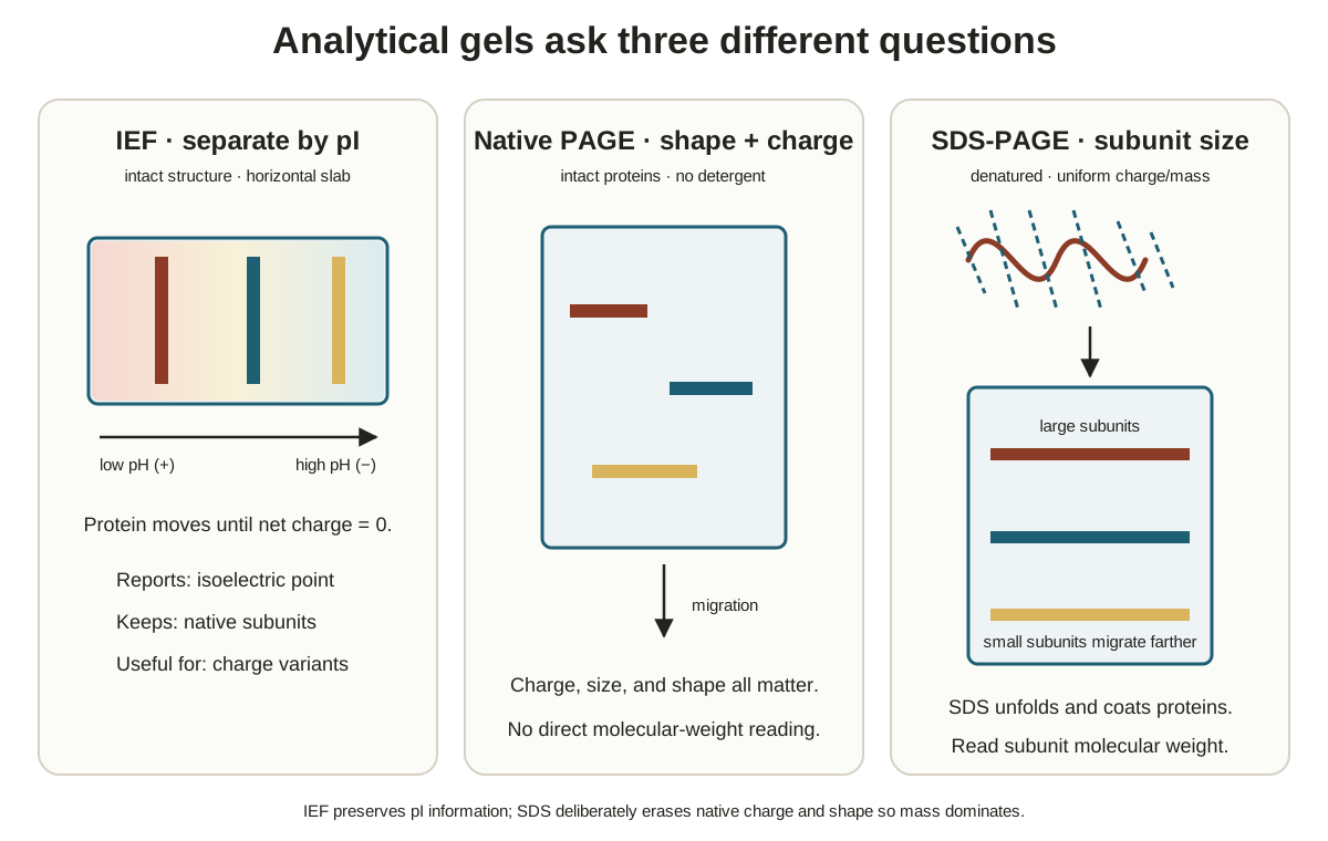
Isoelectric focusing (IEF), native polyacrylamide gel electrophoresis (native PAGE), and the strong-detergent gel introduced later answer different questions because they retain different levels of protein structure and use different separation variables.
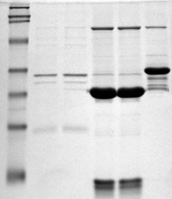
Reference figure · external sourceSDS-PAGE gel with protein bands separated by molecular weight; smaller proteins migrate farther toward the bottom of the gel. Source: Wikimedia Commons · Magnus Manske · CC BY-SA 3.0
Isoelectric focusing and native PAGE
These analytical PAGE gels were described as roughly phone-sized and thinner than a phone.
IEF separates proteins by their pI, defined here as the pH at which a protein’s net charge is zero.
IEF is gentle enough to preserve primary, secondary, tertiary, and quaternary structure.
20 bands establish that at least 20 proteins are present, but a single band may contain several proteins that share an isoelectric point.
IEF can be run in a stable horizontal slab or inside a vertical glass tube. These are alternative geometries for the same separation, and the later two-dimensional method uses the tube format.
Native PAGE is usually run as a vertical slab and separates by both size and charge-to-mass ratio, Q/M.
Smaller proteins tend to move faster.
A larger protein with a sufficiently greater Q/M can move faster than a smaller one, so one native gel cannot reveal which variable caused the faster migration.
“Native” means higher-order structure remains intact. Lactate dehydrogenase (LDH), identified as the last glycolysis enzyme and typically having four subunits, retains those subunits in IEF or native PAGE.
Resolving, stacking, and stopping a slab gel
A vertical slab gel sits between two thin glass plates.
The resolving gel is poured and polymerized first; this is where proteins separate.
The upper gel polymerizes around a Teflon comb.
Removing the comb leaves sample wells in the stacking gel.
A sample well holds about 15 microliters, tops.
Bromophenol blue is commonly added because it does not interact with proteins and moves faster than any protein.
In native PAGE it marks the run front and signals when to turn off the power before proteins leave the gel.
IEF does not require this dye because proteins stop at their isoelectric points; the run is nearly complete when current I falls close to zero.
The stacking gel compresses the initial sample volume into a very thin band before it enters the resolving gel. Without this compression, diffusion in the resolving gel could broaden neighboring protein bands until they appear as one large band.
In response to a student question, the lecturer said stacking requires different buffer and gel pHs plus a lead ion and a trailing ion, which establish a voltage gradient.
A standard example uses a resolving-gel pH of 8.8 and a stacking-gel pH of 6.8.
The trailing ion is slow in the upper gel, accelerates after the pH change, and passes the proteins.
The lecturer corrected the intuitive description: although the region is called a stacking gel, the relevant event is acceleration that produces sharper, more visible bands.
A complete explanation takes about 20 minutes and was deferred to MCB120L.
IEF needs no stacking gel because the electric field itself focuses proteins into sharp bands; after the power is turned off, entropy causes them to spread.
SDS-PAGE removes higher-order structure
SDS was introduced as a man-made strong detergent. Strong detergents carry a full charge such as -1 or +1, whereas mild detergents have polar groups without a full charge.
SDS destroys higher-order protein structure and gives proteins the same Q/M.
SDS-PAGE therefore separates proteins by size alone.
Only primary structure—the linear sequence of amino-acid residues—remains.
Secondary, tertiary, and quaternary structure are lost.
These analytical gels help purify proteins, estimate protein size, detect subunits, and estimate subunit sizes. They can be run in a day or two.
The broader two-dimensional IEF/SDS-PAGE method introduced for later in the lecture instead surveys all proteins in one sample, such as cancer cells, against all proteins in another, such as normal cells.
Gel
Protein state retained
Separation variable
Run interpretation
IEF
Primary through quaternary structure
pI
Proteins focus where net charge is zero; one band can contain several proteins
Native PAGE
Native structure, including subunits
Size and Q/M
Faster movement cannot be assigned to size or Q/M from one gel alone
SDS-PAGE
Primary structure only
Size
Equalized Q/M removes charge as a separation variable
Figure · slide-derivedIEF, native PAGE, and SDS-PAGE preserve different protein features and therefore answer different analytical questions Derived from annotated slide deck 1, p. 21, and annotated slide deck 3, p. 1
Fatty Acids and Lipids as Metabolic Prerequisites
Fatty acids carry substantial energy but would behave as detergents if left free, so cells covalently attach them within storage and membrane lipids.
Fatty-acid shorthand, cis bonds, and energy
Fatty acids are components of lipids rather than a lipid class by themselves.
Saturated fatty acids have no double bonds; unsaturated fatty acids have one or more.
Shorthand
Name stated in lecture
Structural meaning
16:0
Palmitic acid; palmitate when COOH is deprotonated to COO-
16 carbons and no double bonds
18:0
Stearic acid; stearate in the deprotonated form
18 carbons and no double bonds
18:1(9)
Oleic acid
One cis double bond beginning at carbon 9 and extending to carbon 10
18 carbons, two double bonds
Linoleic acid
The second cis double bond begins at carbon 12 and extends to carbon 13
18 carbons, three double bonds
Alpha-linolenic acid
Three cis double bonds
SDS has 12 carbons and a negative charge, while palmitate has 16 carbons and a negative charge. Free fatty acids would therefore act as detergents; high amounts were associated here with sick or dying cells.
Stearate has 18 carbons, whereas glucose has 6. Per molecule, the lecturer said stearate releases much more energy and therefore takes more exercise to burn.
Carbon 1 of a fatty acid is the carboxyl carbon.
For linoleic acid:
the first cis double bond joins carbons 9 and 10
the second joins carbons 12 and 13
the chain continues to carbon 18
“Cis” means that the incoming and outgoing chain portions are on the same side of the double bond, producing a kink.
On an exam, students are usually expected to number fatty-acid carbons to make the drawing easier to grade. Single bonds may be drawn in different rotated orientations, but each specified double bond must remain cis because the enzymes that form those bonds are stereospecific.
More kinked fatty acids let membrane lipids move more easily and affect membrane fluidity.
Exam emphasis: The lecturer introduced this review well before the second midterm, when students will be expected to know the beta-oxidation pathway rather than first encountering fatty-acid structure two days before the exam.
Triacylglycerols store fatty acids and energy
The lecturer said scientists group lipids into seven classes, but he would review only two here.
Acylglycerols use a glycerol backbone, and fatty acids may occupy glycerol carbons 1, 2, and 3.
The three fatty acids can differ.
One possible prompt would place palmitic acid at glycerol carbon 1, alpha-linolenic acid at carbon 2, and another fatty acid at carbon 3.
Triacylglycerols are fats and energy deposits, not membrane constituents. Three long fatty acids plus glycerol provide a large stored-energy source during fasting.
The drawn backbone was identified as L-glycerol because the hydroxyl on carbon 2 was on the left.
The lecturer said research indicates that most organisms use L-glycerol in these compounds.
Archaea use D-glycerol, and he explicitly said, “we don’t know why.”
Unique enzymes attach fatty acids at glycerol positions 1, 2, and 3, and different enzymes remove them from those positions. The lecturer used this to stress that enzymes are fast, restricted, and stereospecific rather than indiscriminate.
He briefly named the convention “stereospecific numbering” and then told students to pretend it had not been introduced. In three dimensions, an active site can use multiple weak bonds—possibly about 20 in his example—to distinguish left- and right-handed molecules even when a paper drawing is confusing.
Phospholipids build membranes
Phosphatidylcholine has:
a glycerol backbone
fatty acids at glycerol carbons 1 and 2
a phosphate at carbon 3
choline attached to that phosphate
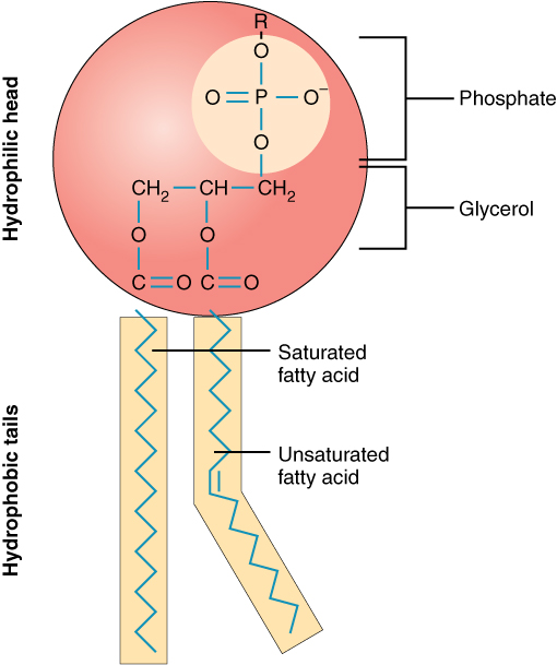
Reference figure · external sourceStructure of a phospholipid showing the glycerol backbone, polar head group with phosphate, and two fatty acid tails including a kink from a cis double bond in an unsaturated chain. Source: Wikimedia Commons · OpenStax · CC BY 4.0
The phosphate forms a diester. The lecturer connected phosphate diesters to the sugar-phosphate backbones of DNA and RNA.
Choline’s nitrogen has four substituents and carries a permanent positive charge at any pH; it is a quaternary amine.
Phospholipids are important because both the plasma membrane and organelle membranes contain them.
Asked whether phosphate can occur only at carbon 3 in the described product, the lecturer answered yes because the placement is enzyme-catalyzed and stereospecific. He first said the enzyme “never makes a mistake,” then qualified the statement: a mutation that caused such a mistake would probably be lethal, so surviving progeny would not be seen.
Replacing choline with the BIS-102 amino acid serine gives phosphatidylserine, illustrating that amino acids have metabolic uses beyond building proteins.
Possible assessment: Draw the phosphatidylserine structure predominant at pH 7, with palmitate at position 1 and stearic acid at position 2.
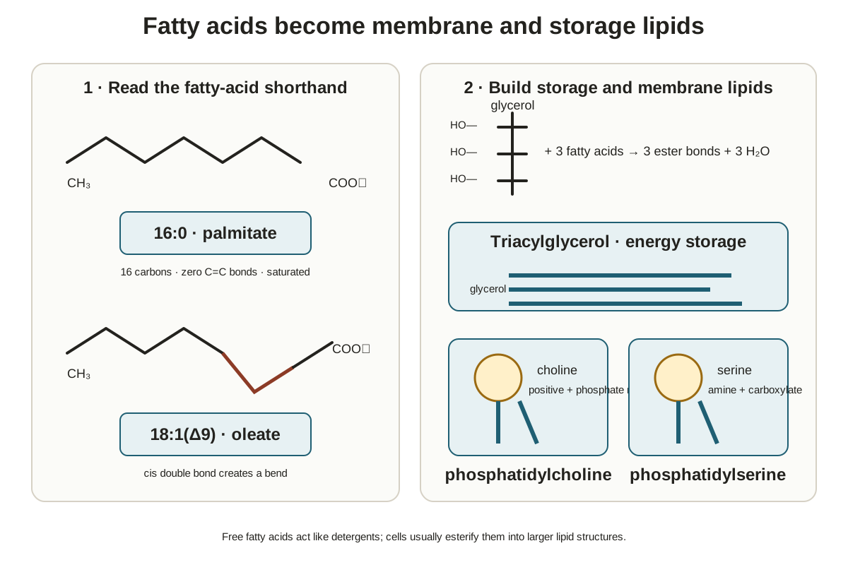
Figure · slide-derivedFatty-acid shorthand and cis geometry connect directly to the glycerol, ester-bond, and head-group logic of storage and membrane lipids Derived from annotated slide deck 1, pp. 22–23, and annotated slide deck 3, pp. 2–3
DNA Microarrays Begin by Isolating Messenger RNA
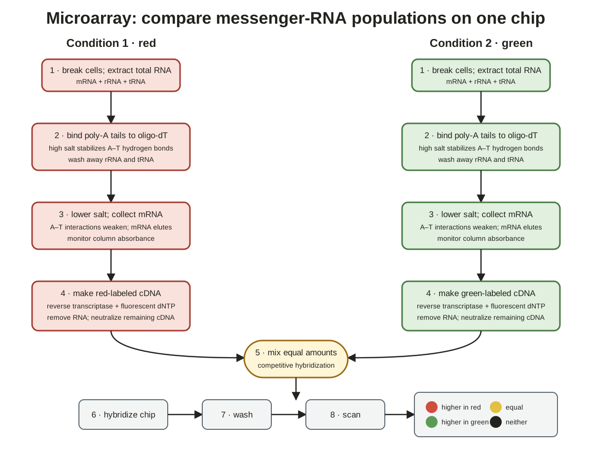
A two-sample DNA-microarray experiment applies the same treatment to cancer and normal cells, then uses poly-A affinity capture to isolate each messenger-RNA population separately.
Matched samples and poly-A capture
DNA microarrays survey all messenger RNAs (mRNAs) made by a cell population.
The lecture’s example compares lab-grown cancer cells with normal cells to ask what differs in cancer.
Organic-solvent extraction yields total RNA, including transfer RNA (tRNA), ribosomal RNA (rRNA), mRNA, and other types. The target population is the mRNA that codes for proteins.
Only mRNA was said here to carry a poly-A tail at its 3-prime end; tRNA and rRNA do not. Nucleic acids are conventionally written 5-prime to 3-prime.
Normal cells are the control. Cancer and normal samples must receive the same procedure on separate, identical affinity columns.
The isolation proceeds in source order:
Fill each small column with polar, highly porous microscopic beads held above a fritted plastic support.
The frit’s small holes pass buffer and dissolved material but retain the beads.
The packed bead bed is the affinity-column resin, and tubing or a valve controls the flow rate.
Covalently attach polydeoxythymidine to the beads as the ligand.
No arm is needed because the target mRNAs are linear rather than three-dimensional enzyme active sites.
Load the total-RNA population.
Polydeoxythymidine acts as a “fish hook” for the mRNA poly-A tail.
mRNA binds near the top, while tRNA and rRNA flow through.
A–T contacts continually break and reform, sometimes at a neighboring ligand; capture is dynamic and noncovalent.
Collect fractions and monitor absorbance at 260 with a spectrophotometer.
RNA bases contain conjugated double bonds that absorb ultraviolet light.
The lecturer imagined collecting perhaps 100 Eppendorf tubes.
Early absorbance is high because unwanted tRNA and rRNA are leaving the column.
Wash with buffer until the absorbance approaches zero while mRNA remains bound.
The pH should maximize binding.
Perhaps pH 7.5 was offered as a trial-and-error or literature example, not a fixed value.
Elute the mRNA population.
A large shift to either very high or very low pH can disrupt hydrogen bonding.
Alternatively, lowering the salt concentration releases the mRNA.
Either method should produce an elution peak containing the mRNA population.
The unwanted early fractions are the flow-through.
A buzzer marked a five-minute break. Before pausing, the lecturer said the first point after the break would be why low salt causes release.
Why lower salt releases the mRNA
Lowering cation concentration exposes repulsion between negatively charged nucleic-acid backbones, allowing the captured strands to separate.
After the break, the lecturer first restated that separate columns had yielded cancer-cell and normal-cell mRNAs, then returned to the deferred mechanism.
A nucleic-acid duplex carries negative charges along both phosphodiester backbones; at the experimental pHs the phosphate groups were represented as PO-.
Cations such as magnesium or zinc reduce this repulsion. In cells, positively charged arginine and lysine residues in proteins can also do so.
A buffer containing fewer cations no longer tones down the backbone repulsion, so the strands separate and the mRNA leaves its ligand.
Reverse Transcription Produces Two Colored DNA-Copy Pools
Each isolated mRNA population is copied into fluorescent complementary DNA (cDNA), freed from its RNA template, and returned to pH 7 before chip binding.
Copy, hydrolyze, and neutralize
Add excess oligo-dT primers to one isolated mRNA pool.
A pool contains hundreds or thousands of distinct mRNAs.
Polymerases require a double-stranded starting point, so the primers bind poly-A tails and give every mRNA a primed end.
Add reverse transcriptase and deoxynucleoside triphosphates.
Reverse transcriptase uses each RNA as a template for a single DNA strand.
The triphosphates supply energy for covalent-bond formation, while the enzyme catalyzes polymerization.
Fluorescently modify one nucleotide type.
The example arbitrarily labels deoxy-ATP red.
Excitation produces emission at a longer wavelength and therefore a color.
Labeled deoxy-ATP is incorporated occasionally opposite transcript uridine; not every A must be labeled, but a fair number must be.
Complete the red cDNA population.
The procedure converts colorless mRNA information into a colored DNA copy.
DNA was described as more stable than RNA.
The lecturer said the c in cDNA means copy.
Destroy the RNA template so the cDNA becomes single-stranded.
Incubate in 1 molar NaOH, heat, and allow 15 minutes.
Hydroxide removes the proton from RNA’s 2-prime hydroxyl, creating O-.
Electronegative oxygens leave phosphorus partially positive; the nearby O- attacks phosphorus and breaks a bond by substitution.
This process breaks covalent bonds throughout the mRNA, while DNA lacks the 2-prime hydroxyl and was described as stable under the treatment.
Neutralize after 15 minutes by adding HCl to restore pH 7.
This step is critical because the high-pH cDNA cannot form the hydrogen bonds needed to bind the chip.
Repeat the procedure for the second sample with a green fluorescent nucleotide.
Cancer-cell mRNAs become red cDNAs.
Normal-cell mRNAs become green cDNAs.
Green is deliberately chosen for the comparison, although another color could be used.
A student clarified that the question was why deoxy-ATP rather than deoxy-GTP was labeled. The lecturer corrected his initial interpretation and answered that the choice is arbitrary: any one nucleoside triphosphate could be red, but labeling all of them would be too expensive.
Competitive Hybridization Converts Binding into a Color Map
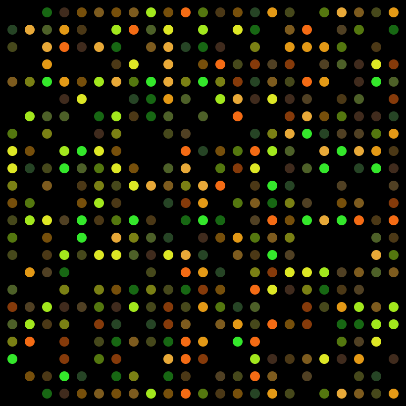
Red cancer cDNAs and green normal cDNAs compete for a limited number of complementary gene-specific sites on one chip, and their relative binding determines each spot’s color.
Reference figure · external sourceDiagram of a DNA microarray (gene chip) with a grid of probe spots used to detect gene expression via competitive hybridization of fluorescently labeled cDNA. Source: Wikimedia Commons · Guillaume Paumier · CC BY-SA 3.0
Chip construction, hybridization, and scanning
The overall procedure is expensive because the affinity columns, purified reverse transcriptase, and other components cost money.
A microscope-slide-sized device was said to cost about $800.
Its chip contains machine-deposited spots of single-stranded DNA, each approximately one nanoliter.
Each spot contains several identical copies from one gene.
Neighboring spots represent different genes.
Spots should have about the same copy number.
A chip might represent one human chromosome.
The DNA spots act as receptors.
The company knows which gene is at each position and what it codes for because it isolated, copied, and positioned the DNA. The customer is principally paying for this identity information.
The two-color comparison then proceeds:
Place the slide in a small container with the company-optimized buffer, pH, incubation time, and temperature.
Add a small portion of red cancer cDNA and a small portion of green normal cDNA.
The source tubes are not exhausted, so replicate experiments remain possible.
Rotate the container during incubation to mix the solution.
Allow red and green cDNAs to compete for the limited complementary sites.
Rinse the slide to remove nonspecific binding to the glass.
Scan each spot separately for red and green fluorescence, then merge the images.
Figure · slide-derivedThe two samples remain separate through labeled cDNA production, then compete together on one chip before channel scanning and color interpretation Derived from annotated slide deck 1, pp. 26–31, and annotated slide deck 3, pp. 4–9
Four outcomes and their limits
Spot color
Binding outcome stated in lecture
Red
More cancer-derived cDNA bound than normal-derived cDNA
Green
More normal-derived cDNA bound than cancer-derived cDNA
Yellow
Approximately equal red and green binding; the gene is expressed in both compared conditions
Black
Nothing bound
In the yeast example, green marked genes expressed during normal growth, while red marked genes expressed as yeast began forming spores.
Yellow spots were detected in both conditions.
Green and red spots were skewed toward one condition.
Many spots were black because not every gene is active, and different cell types express different protein sets.
In response to an unrecoverable student question, the lecturer’s guess—and explicitly “just a guess”—was that more sophisticated instruments provide numerical values for everything. The class would use only the overall color picture.
Red spots would be of special interest in the hypothetical cancer comparison, but a red gene need not cause cancer.
One affected regulatory protein could influence about 20 other spots.
The method surveys all genes at once, unlike isolating one target at a time, but substantial follow-up remains necessary.
The technique is powerful but not perfect; the lecturer said it “can be lied to.”
Homework 1 includes a question directing students to discuss the technique online.
Companies try to make every spot the same, but the lecturer said a normal distribution means some may contain slightly more or less material. The expected result still contains many different colors.
First-Dimension IEF Surveys the Proteome
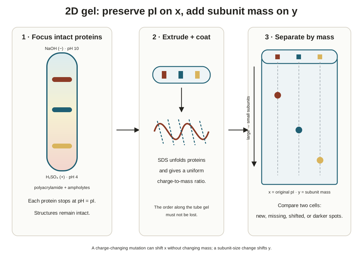
Two-dimensional IEF/SDS-PAGE examines all proteins—the proteome—by focusing matched samples across the same broad pH range before any size separation.
A single-base substitution may escape microarray detection because the remainder of the polymer can still bind.
If the substitution changes charge, IEF and native PAGE can reveal a difference. SDS-PAGE will not reveal that charge difference because SDS coats the proteins.
Polymerizing a tube gel
Stopper or seal one end of a glass tube.
Add acrylamide and bisacrylamide.
Bisacrylamide makes cross-links.
Without it, the mixture remains watery instead of forming a gel.
Add small organic molecules rendered in the transcript as amphilites.
Under an electric field, they form the pH gradient.
Add TEMID, which the lecturer said prolongs the lifetime of free radicals.
Add ammonium persulfate as the free-radical source.
Its unstable oxygen–oxygen bond breaks in water.
Each sulfate fragment receives one unpaired electron.
The radicals initiate a chain polymerization that builds the gel network.
Without ammonium persulfate, no gel forms; without TEMID, the radicals do not persist long enough to drive the chain reaction, so no gel forms.
Allow about 15 minutes for a clear gel to form throughout the stoppered tube.
Establishing the gradient and focusing matched samples
Place 60 millimolar NaOH in the upper reservoir next to the negative electrode.
Place 60 millimolar sulfuric acid in the lower reservoir next to the positive electrode.
These high- and low-pH extremes trap the small pH-gradient molecules within the gel.
To survey all proteins, the lecturer proposed a broad pH 2 to pH 10 gradient rather than a narrow pH 3-to-4 interval.
The molecules are purchased for a desired range. The lecturer estimated that perhaps 1 mL costs about $50, reflecting the cost of purification.
Apply a protein extract and turn on the power.
The illustrative red color on the slide did not mean the proteins themselves were colored; proteins usually have no color.
A whole-proteome run could form a hundred bands, and multiple proteins may share one pI.
This first dimension separates proteins by pI.
Extract cancer-cell and normal-cell proteins separately and focus them in different tubes across the same pH 2-to-10 range.
The apparatus can run about 10 or so tubes.
Proteins remain structurally intact after IEF.
SDS Treatment and the Second Dimension Separate by Size
After IEF fixes each protein’s horizontal pI position, SDS denatures the proteins and equalizes Q/M so the vertical slab gel separates subunits by size alone.
Denaturing and coating the focused proteins
Extrude each tube gel with a plunger into a tray of SDS, like pushing out a thin piece of spaghetti.
Cancer and normal gels receive the same treatment.
Small SDS molecules diffuse into the gel, while the proteins are too large to diffuse out.
Use excess SDS to disrupt protein structure.
SDS is a 12-carbon, man-made compound.
Its 12-carbon tail is nonpolar, and its head carries -1.
The full charge makes it a strong detergent suited to disrupting proteins.
The carbon chain rotates in solution rather than remaining straight.
Add excess mercaptoethanol to reduce disulfide bonds.
Most proteins were said not to contain internal disulfides.
Oxygen entering during laboratory work can create artificial disulfides within or between subunits and distort the result.
Heat the sample to accelerate denaturation.
Allow SDS to coat the unfolded subunits.
Hydrophobic tails bind hydrophobic amino-acid side chains; leucine, isoleucine, and tryptophan were examples.
Negatively charged heads face the surrounding water.
Gaps remain at polar or charged residues; glutamate and arginine were examples.
Equalize charge-to-mass ratio.
Thousands of SDS molecules coat an unfolded subunit, leaving primary structure but no biological activity, alpha helices, beta sheets, tertiary structure, or intact quaternary structure.
A larger subunit binds proportionally more SDS than a smaller one, giving both the same Q/M.
An example initial charge of +8 becomes negligible beside thousands of bound negative charges.
The coated subunits repel one another; the lecturer called them clustered micelles.
Running the one-wide-well SDS-PAGE slab
Pour and polymerize the resolving gel.
Pour the stacking gel around a Teflon comb that creates one giant indentation rather than many small wells.
Place the entire SDS-soaked IEF tube gel into that wide well.
SDS is present throughout.
Typical/example concentrations were about 4% stacking gel and 10% resolving gel, although the lecturer said the choices can vary.
The gel pHs serve a particular reason that was not developed here.
Put the positive electrode below the gel and the negative electrode above it.
Exam emphasis: Electrode polarity was presented as likely first-midterm material. SDS-coated proteins are strongly negative, so a negative lower electrode would drive them upward and out of the separation gel.
Turn on the power.
Proteins first stack into thin bands and then move downward.
The first dimension separated by isoelectric point.
The second dimension separates by size, with smaller proteins moving fastest.
Add bromophenol blue with some glycerol above the gel and follow its run front.
The front may be slightly curved because salt concentration and electric field are not perfectly uniform.
Turn off the power before the dye runs off, or proteins may also leave the gel and invalidate the observed population.
Remove one glass plate and roll the gel into stain.
The proteins are already denatured, so further loss of activity from staining is irrelevant.
The second dimension can reveal hundreds, if not thousands, of spots.
Run cancer and normal samples on separate gels, then merge their images to find differences.
Figure · slide-derivedThe first dimension fixes each protein’s pI position; SDS treatment then preserves that x-position while subunit mass determines the second-dimension y-position Derived from annotated slide deck 1, pp. 32–36, and annotated slide deck 3, pp. 10–14
Reading Two-Dimensional Spot Maps and the Lecture Boundary
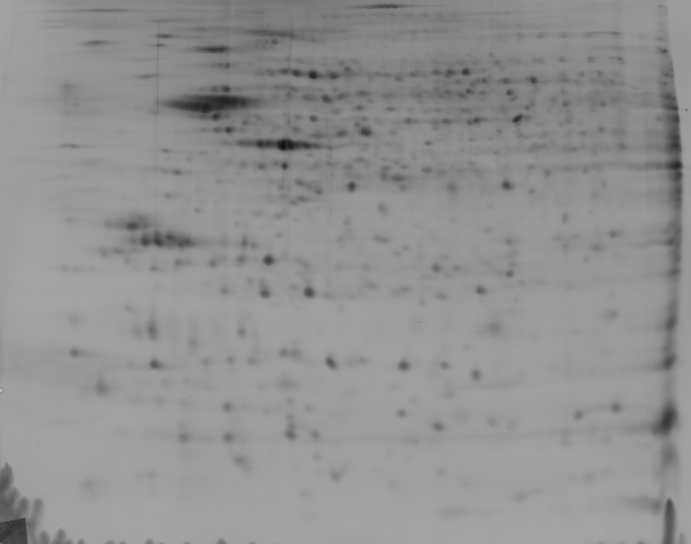
A two-dimensional spot map is read by pI along one dimension and molecular weight or size along the other, so aligned spots reveal shared separation properties.
Reference figure · external source2D gel electrophoresis of rat hippocampal proteins separated by isoelectric point (pH 4-7, horizontal) and molecular weight (vertical), showing the characteristic spot pattern used to map a proteome. Source: Wikimedia Commons · Jean-Etienne Minh-Duy Poirrier · CC BY-SA 2.0
Radioisotopes were described as more sensitive than staining.
In the example, the lecturer said the organism was probably grown with S35 methionine.
After IEF and SDS separation, film was placed over the gel in a freezer for a week or so and then developed to reveal proteins.
In the E. coli example:
spots on the same vertical line share a pI
a different vertical line represents a different shared pI
spots on the same horizontal line share molecular weight or size
spots lower on the gel are smaller
The lecturer said the class had covered the equivalent of four lectures in a normal quarter and was already beginning normal-quarter week two.
Microcalorimetry was not taught in this session. The lecturer had brought a light for it but ran out of time and deferred this third and final overall technique to first thing the next day.
Study Review Questions
Why must an affinity-column bead used in this lecture be porous without acting as a molecular sieve?
How do IEF, native PAGE, and SDS-PAGE differ in the protein structure they preserve and the variable or variables by which they separate proteins?
How do the lead ion, trailing ion, and pH change sharpen proteins before they enter a resolving gel?
Why are free fatty acids detergent-like, and how do triacylglycerols and phospholipids place fatty acids into different cellular roles?
How does a polydeoxythymidine affinity column isolate mRNA from total RNA, and why does lowering salt release the captured mRNA?
What ordered steps convert isolated mRNA into a fluorescent, single-stranded cDNA population that can bind a microarray chip?
What do red, green, yellow, and black microarray spots mean in the cancer-versus-normal comparison?
Why can a microarray miss a single-base substitution that a charge-sensitive protein separation might reveal?
How do ammonium persulfate and TEMID contribute to tube-gel formation, and how is the broad pH gradient established?
Why must the lower electrode be positive in the second dimension, and how are vertical and horizontal alignments interpreted on the final spot map?



.jpg)