The lecture introduced BIS 103 as a study of intermediary metabolism, with emphasis on pH, enzymes, energetics, and the connections among pathways. It framed animal, plant, and bacterial cells as dynamic chemical systems and introduced three complementary ways to compare whole-cell metabolism. The second half refreshed buffer behavior, the lactate dehydrogenase reaction, phosphate-buffer preparation, and enzyme kinetics. The lecture ended while beginning affinity chromatography, with the construction of the affinity matrix left unfinished until the next class.
Course opening, expectations, and assessment
Dr. Hilt opened BIS 103 by establishing the people, tools, and working expectations for a compressed six-week course.
Learning assistants Cat and Wiley previously took BIS 103 and hold office hours. They receive one unit of MCB 197T for three hours of work per week.
Students interested in being learning assistants during the coming academic year may email klhilt@ucdavis.edu. Dr. Hilt said he would not teach BIS 103 that academic year but would teach two large BIS 105 sections in winter; he described BIS 105 as half BIS 102 and half BIS 103.
The only required book is a practical workbook. Unlike larger biochemistry texts, it is intended to help with calculations that are useful in a research laboratory.
The course studies intermediary metabolism, the biochemical reactions in living cells. These reactions depend strongly on pH, and cells use buffers.
pH affects hydrogen bonds and ionic bonds.
Experimental biochemistry requires recognizing charges and hydrogen bonds on biomolecules, preparing buffers, and reasoning about charge.
Metabolism works because of enzymes. Dr. Hilt estimated that 99.99% of cellular steps are enzyme-catalyzed and said most enzymes are proteins.
Cells continually spend energy from consumed material to build proteins, DNA, cell walls, and other structures. A major course goal is to understand how cells capture and use that energy, while recognizing that science does not yet explain everything.
Students were asked to meet nearby classmates because discussing and arguing through concepts is important.
The ten-week course is compressed into six weeks and meets Monday, Tuesday, and Wednesday.
There are two midterms and a final.
The first midterm is moved to week three, after about two and a half weeks of meetings, which concentrates exams near the end.
Summer session has no separate final-exam week; the final occurs on the last class day.
Room and accommodation arrangements were still uncertain. Dr. Hilt said he would try to obtain an extra exam room and find out whether accommodated exam services were available under the current budget.
Each midterm lasts 50 minutes, with no lecture afterward. The final lasts 1 hour 50 minutes, although Dr. Hilt expected most students to finish in about an hour.
Exam questions include calculations and short answers, with occasional multiple-choice questions and no Scantron. Students must bring a calculator.
Equations and constants for weak acids and bases, enzyme kinetics, energetics, and similar calculations are provided. Students must know how to use them rather than memorize them.
Exams cover lecture and homework. Only Homework 1 applies to the first midterm; it is ungraded but strongly emphasized and was to be posted the next day.
The final is worth 200 points, twice a midterm.
About half acts like a third midterm on material after the second-midterm boundary.
The other half revisits material that should have been or was tested on Midterms 1 and 2.
Dr. Hilt noted that placing the boundary about a week before the final is difficult in a three-meeting week.
The course is curved: whatever the average is becomes the lowest B−, and roughly the top 15% receive some form of A. Dr. Hilt described the tests as challenging and requiring hard work, while saying he wants students to pass.
The big picture of metabolism and its recurring themes
The course will isolate pathways for study without losing sight of the fact that metabolism is a connected, regulated whole.
Metabolism includes all reactions in a cell, more than the course can cover. Glycolysis will be studied separately, but it remains connected to other pathways.
Three techniques for obtaining a larger view of cellular metabolism were previewed for later in the lecture.
The selected pathways are ones biochemists generally need to know. Esoteric pathways and the often-complicated amino-acid biosynthetic pathways are left for other study.
For each selected pathway, students must understand what it does, recognize that it may serve more than one function, work with its enzymes, and relate it to other pathways.
An assay is a particular way to measure something. An enzyme assay measures change, specifically product made per unit time.
Every step in a pathway such as glycolysis is enzyme-catalyzed, and enzyme activity must be controlled.
Inhibition slows conversion when less is needed.
Activation speeds conversion when more is needed.
Enzymes catalyzing reactions with very large negative ΔG values especially require control because those reactions are thermodynamically spontaneous in the lecturer's framing.
Dr. Hilt strongly recommended MCB 120L, a lecture/laboratory course with labs on the third floor of ESAU. He said laboratory mistakes and arguments with partners reinforce BIS 102/103 concepts and can improve preparation for the MCAT and professional-school goals.
Four types of weak bonds from BIS 102 recur throughout the course. They are noncovalent interactions that can form and release.
Multiple weak bonds in an enzyme active site allow recognition of the correct substrate.
Weak bonds also support protein structure, membranes, and double-stranded DNA.
Exam emphasis: Students must know glycolysis “like the back of your hand” for the first midterm. It appears on the last page of Homework 1 even though step-by-step instruction may not begin until the end of that week or the beginning of the next; names and structures can be learned before the chemistry and cellular purpose are explained.
Change in entropy, ΔS, is another recurring concept.
Some enzymes require a specific metal ion at the active site in addition to their amino-acid structure.
Examples included Fe²⁺ and Mg²⁺.
For an enzyme that requires Mg²⁺, replacing it with Mn²⁺ or Cu²⁺ does not work because the atomic orbitals differ.
Some proteins position two particular, sometimes different, metals next to each other so that they act together in the required chemistry.
Only small dietary quantities of metals are needed, and a normal diet supplies plenty.
Proton gradients across membranes are another course theme.
Moving positive charge to create more protons on one side of a membrane requires energy.
In eukaryotic mitochondria, the electron transport chain creates a critical proton gradient.
In chloroplasts, light reactions involving photosystems 1 and 2 also contribute to proton gradients.
Dr. Hilt said these processes account for most ATP production across organisms.
The course will examine mitochondrial proton gradients deeply in its latter third and photosynthesis near the end. Fatty-acid biosynthesis was only a possible final topic if time and capacity allowed.
Cells as open chemical engines
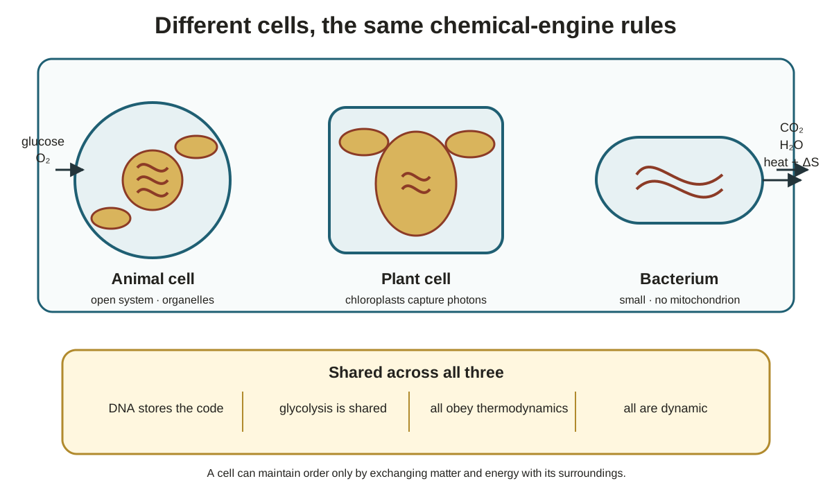
An animal cell survives as an open chemical engine by taking in matter, exporting waste, and capturing only part of the available energy.
A generalized animal cell takes in nutrients such as glucose and oxygen to maintain order and conduct needed reactions.
It expels waste rather than accumulating it.
CO₂ is a major breakdown waste product, which is why people exhale it.
Water is another product compatible with cells.
Some energy is always lost as heat or entropy because a cell cannot capture all nutrient energy.
The cell obeys thermodynamics.
ΔG is free energy available for work such as movement, division, and biosynthesis.
ΔH is change in heat or enthalpy.
T is temperature in kelvin.
ΔS is entropy or disorder.
Converting one ordered six-carbon glucose into six separate CO₂ molecules illustrates increased disorder.
Biochemical standard free energy, ΔG′, provides a human standard for comparing reactions.
Every reaction component is at 1 M except protons.
Unless stated otherwise, the prime convention uses pH 7, so [H⁺] = 10⁻⁷ M. A 1 M proton concentration would be pH 0 and incompatible with most cells, although Dr. Hilt noted that some organisms tolerate pH 0.
Glucose breakdown to CO₂ and water has ΔG′ = −686 kcal/mol. The number is not a memorization target; its large magnitude is the point.
ATP synthesis from ADP plus inorganic phosphate (Pᵢ) has ΔG′ = +7.2 kcal/mol, so energy must be supplied.
The large energy release from glucose breakdown is thermodynamically sufficient for making many ATP molecules.
The course uses kcal/mol rather than joules because the workbook, written in 1979, uses kcal/mol; conversion by a constant is possible.
Cells continuously take in multiple nutrients, produce waste, capture some energy in ATP, and perform other work. Their components are dynamic: mitochondria move, fuse, and divide.
Systems biology describes the idea that everything in a cell communicates with something else. Changing cellular ATP concentration would affect many processes, and pathways occur simultaneously even when studied one at a time.
Dr. Hilt said biology is only beginning to address quantum mechanics or quantum biology. Protons, electrons, and photons behave unlike objects in everyday experience and remain active in cells.
He explicitly cautioned that intermediary metabolism is complex and incompletely known; the course will describe what is known without claiming complete understanding.
Plant chloroplasts receive photons, and photosynthesis will be the course's last major topic, where some earlier ideas recur.
Bacteria such as E. coli are small enough that the shown magnification requires electron microscopy. They are long-used biochemical models; a human DNA fragment or gene can be put into a microbe in hopes that it will produce many copies of the encoded protein for study.
Animal, plant, and bacterial cells share DNA as their code, pathways such as glycolysis, thermodynamic laws, dynamic behavior, and complexity.
The lecturer said students would know glycolysis well by “August 19th or whatever” date the syllabus gives, not as a firm date.
Research makes scientists “pretty sure,” in Dr. Hilt's wording, that mitochondria and chloroplasts evolved from bacteria.
Figure · slide-derivedDifferent cell types share the same chemical-engine constraints while exchanging matter and energy with their surroundings Derived from annotated slide decks 1 and 2, pp. 5–8
From pathway chemistry to enzyme behavior and applications
Knowing that one compound becomes another is only the start; a biochemist must also understand the enzyme, rate, energetics, measurement, and purpose of the reaction.
In glycolysis's first step, glucose becomes glucose 6-phosphate. Students will eventually need to draw glucose with phosphate on carbon 6.
At a cell's operating temperature—37 °C in the human example—uncatalyzed reactions are not fast enough for survival.
Every chemical step therefore needs an enzyme.
Enzymes are almost always proteins, with unspecified exceptions.
Kinetics asks how fast a reaction occurs. Thermodynamics asks whether it has the potential to occur.
Even a reaction with a large negative ΔG has no kinetic chance in the cell without an enzyme.
Enzyme measurements characterize different aspects of behavior.
Vmax asks how fast an enzyme can operate once substrate is bound.
Km, the Michaelis constant, is presented as the usual measure of how well an enzyme binds substrate.
Turnover number is the number of substrate molecules converted to product by one enzyme molecule in one minute. Dr. Hilt gave about 30,000 per minute as typical for most enzymes, helping explain why a cell need not make many copies of an effective enzyme.
Kᵢ is the equilibrium dissociation constant of inhibitor I. An inhibitor binds an enzyme and slows it, providing one form of control.
Enzyme assays can use spectrophotometry, radioisotopes, or fluorescence.
A radioactive substrate or product can make enzyme activity detectable.
If the target enzyme's substrates do not absorb light, other enzymes and compounds can couple its reaction to a light-absorbing readout.
Enzyme mechanism asks which active-site chemical groups perform the chemistry that converts substrate to product.
Mechanistic knowledge may later support compounds that inhibit an enzyme implicated in cancer.
Dr. Hilt broadly framed disease as disrupted metabolism involving something wrong with one or more enzymes; treatment may seek to eliminate or reduce an enzyme's activity.
Biochemistry tracks change in free energy, enthalpy, entropy, and ΔE.
ΔE applies to redox reactions.
Dr. Hilt said most metabolic reactions studied in the course involve electron flow: one compound donates electrons and another accepts them. A concrete ΔE example was deferred.
Reasons to study metabolism include medicine, fundamental research with possible later benefits, and crop improvement.
One proposed application is adding one or more genes to a crop so that it can convert atmospheric nitrogen directly, especially where fertilizer is unaffordable.
Comparing organisms can reveal pathway relationships, and microbes or cultured human cells can enable discoveries when direct work with humans is not possible.
Metabolism was summarized as the chemistry of life.
At 1:00, halfway through the meeting, the class took a five-minute break. Students could ask Dr. Hilt questions before lecture, after lecture, or during the break.
Three complementary views of whole-cell metabolism
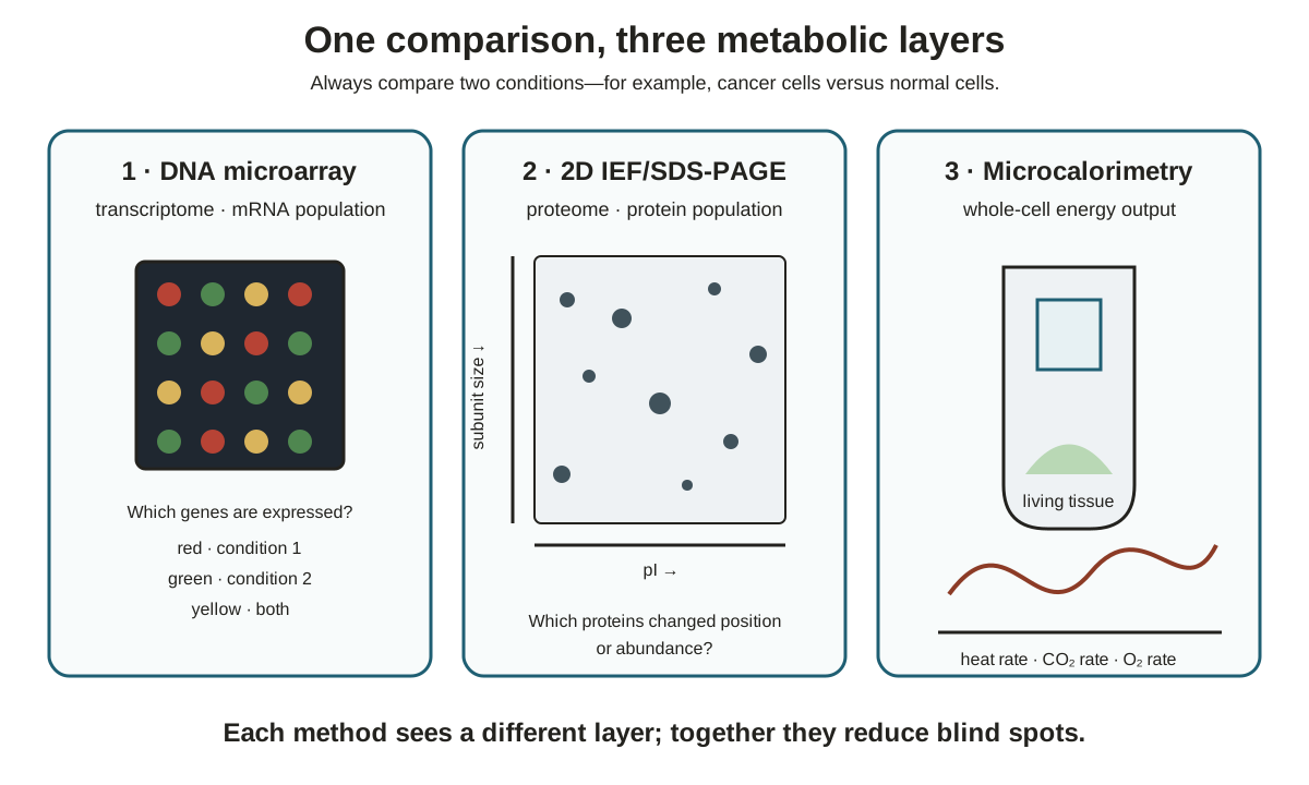
DNA microarrays, two-dimensional gels, and microcalorimetry compare cells at different measurement layers, so no single imperfect technique has to carry the whole conclusion.
Homework 1 was to include an online DNA-microarray activity.
The example throughout compared cancer cells with normal control cells.
DNA microarrays
DNA microarrays ask which messenger RNAs (mRNAs) a cell is making at the time of investigation.
All mRNAs coding for proteins are extracted and compared, with many differences usually expected between cancer and normal cells.
Looking across those mRNAs is studying the transcriptome, one whole-cell view of metabolism.
Two-dimensional gels
Isoelectric focusing (IEF) separates proteins by isoelectric point (pI) in one direction.
An SDS-PAGE gel runs in a second direction and separates proteins into subunits.
Comparing the resulting protein patterns surveys the proteome, the proteins being made.
The method can complement a microarray. An mRNA base-pair change might escape the microarray readout, but a protein change from an uncharged valine R group to a charged glutamate R group can appear on a two-dimensional gel.
No scientific technique is perfect, so a subject should be measured from multiple perspectives.
Microcalorimetry
Microcalorimetry uses an instrument sensitive to very small amounts or flows of heat released by living material.
With an appropriate setup, the same instrument can also yield:
rate of CO₂ production, such as CO₂ produced per minute;
oxygen consumption, which measures mitochondrial function in eukaryotic cells because most inhaled oxygen goes to mitochondria.
Whether cancer cells produce CO₂ at a higher or lower rate than normal cells is unknown until it is measured.
Figure · slide-derivedThe transcriptome, proteome, and whole-cell energy readouts answer complementary versions of the same comparative question Derived from annotated slide decks 1 and 2, p. 11
Mitochondrial and chloroplast biochemistry ahead
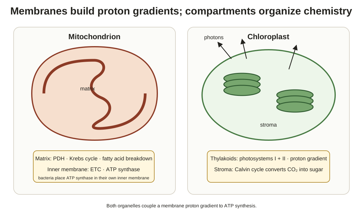
The course's last third will concentrate on the compartments and membrane machinery that connect carbon metabolism to ATP production.
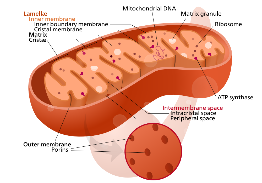
Reference figure · external sourceMitochondrion internal structure, showing the matrix where the Krebs cycle and fatty acid breakdown occur and the inner membrane where the electron transport chain and ATP synthase are embedded. Source: Wikimedia Commons · Kelvinsong (modified by Sowlos, IsadoraofIbiza) · CC BY-SA 3.0
In mitochondria, the course will examine:
the pyruvate dehydrogenase (PDH) complex;
matrix enzymes of the Krebs cycle;
fatty-acid breakdown in the matrix;
thousands of electron transport chains (ETCs) embedded in the inner membrane;
ATP synthase embedded in that membrane.
Dr. Hilt described ATP synthase as making most ATP on the planet. Bacteria lack mitochondria but have ATP synthases in their inner membrane.
In chloroplasts:
light reactions involving photosystems 1 and 2 occur in thylakoid membranes;
the surrounding liquid is the stroma;
enzyme-catalyzed Calvin-cycle reactions in the stroma convert CO₂ into sugar.
Dr. Hilt again said the biochemistry of these organelles is not completely known.
This organelle preview closed the first slide set. The second set refreshed selected BIS 102 material needed for metabolism: weak acids and bases, enzymes in general, and lipids.
Figure · slide-derivedMembranes house proton-gradient machinery while the surrounding compartments organize carbon metabolism Derived from annotated slide decks 1 and 2, pp. 12–13
Designing a buffer for a biological experiment
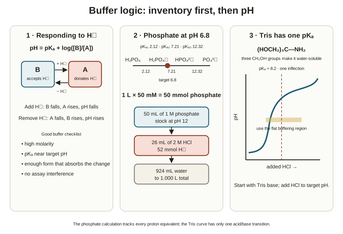
A useful buffer must have enough material to absorb a proton disturbance, a suitable pKa, and no unwanted effect on the enzyme or assay.
A cellular or laboratory buffer needs high molarity because one or two molecules cannot control pH.
For a cell extract, Dr. Hilt gave 50 mM or higher as workable.
A 1 µM buffer will not work.
A buffer is usually chosen with a pKa near the desired pH; pH 7.5 was the example target.
If the studied reaction produces protons, the starting buffer should contain more weak base B than acid A.
B accepts incoming protons.
Protonated A cannot accept another proton.
The buffer should control pH without speeding or slowing the enzyme and without interfering with the assay. In a spectrophotometric assay, it must not absorb at the measurement wavelength.
Henderson–Hasselbalch proton accounting
The Henderson–Hasselbalch equation relates pH to pKa and log([base]/[acid]).
When H⁺ is added, B decreases by the amount added and A increases by that amount because B + H⁺ becomes A.
Excess acid can exhaust B, after which pH falls sharply; buffer capacity is limited.
Removing protons has the opposite effect and raises pH.
A proton may leave by transport across a membrane to create a pH gradient.
A chemical reaction may incorporate the proton into a compound.
Lactate dehydrogenase links proton balance to metabolism
The lactate dehydrogenase reaction directly connects buffer behavior, proton consumption, redox chemistry, and the final step in the lecture's glycolysis scheme.
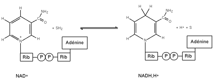
Nicotinamide adenine dinucleotide (NAD) appears as reduced NADH and oxidized NAD⁺.
Pyruvate has three carbons, with its carboxyl group at carbon 1.
The carboxyl pKa is 2.5 and is not a memorization target.
The course usually considers cells near pH 7 ± 1, far above that pKa, so the deprotonated base form predominates.
When pH equals pKa, acid and base are 50:50.
Students are expected to learn the predominant structures at pH 7, including why the molecule is called pyruvate rather than pyruvic acid.
Hydrogen states and hydride carriage
NADH carries two electrons and a proton as a hydride.
Dr. Hilt distinguished three hydrogen states:
H⁺ has no electron in the stated comparison;
H· has one electron and no charge;
H⁻ is hydride with two electrons.
Reference figure · external sourceRedox interconversion of NAD⁺ and NADH, showing the nicotinamide ring that accepts a hydride (H⁻) during reduction. Source: Wikimedia Commons · NicolasGrandjean · CC BY-SA 3.0
NADH participates in multiple pathways and safely carries hydride energy without releasing it merely by colliding with membranes, proteins, or DNA.
An enzyme such as LDH can unlock that energy when NADH binds at its active site.
Lactate has a hydroxyl group on carbon 2. LDH is stereospecific and produces L-lactate, drawn with the OH on the left, rather than alternating between L- and D-lactate.
In the forward direction, the reaction consumes a proton by incorporating it into lactate.
Without buffer, pH would rise.
Excessive pH rise could unfold and inactivate the enzyme.
This reaction is the last step of the glycolysis scheme used on Homework 1 and is reversible.
Exam emphasis: For glycolysis, students must draw structures, know enzyme names, and know whether each reaction is reversible.
Worked phosphate-buffer preparation
Preparing 1 L of 50 mM phosphate buffer at pH 6.8 from a 1 M phosphate stock at pH 12 requires both concentration accounting and two protonation-state calculations.
Available titrants are 2 M HCl and 3 M NaOH.
Calculation workflow
Set the phosphate amount. Use 50 mL of the 1 M phosphate stock. The other ingredients contain no phosphate, and most of the final liter will be water.
Choose the titrant direction. Phosphoric acid is polyprotic, with three protons and three pKa values, so all species should be written out during an exam. At pH 12, near the highest pKa, the stock contains some PO₄³⁻ and more HPO₄²⁻. Reaching pH 6.8 requires HCl; NaOH moves pH in the wrong direction.
Find the starting base fraction. The first Henderson–Hasselbalch calculation gives a base/acid ratio of 0.4786.
Interpret this as 0.4786 base units per 1 acid unit, or 0.4786 out of 1.4786 total units.
The result is 32% base.
If pH equaled the relevant pKa of 12.32, the mixture would be 50:50. Because pH 12 is below that pKa, less than 50% base is a reasonableness check.
Find the final acid fraction. At final pH 6.8, use the relevant pKa of 7.21 and solve for acid.
Acid is 1 out of 1.389 total units, or 72%.
Because pH 6.8 is below pKa 7.21, more than 50% acid is a reasonableness check.
Find the proton requirement. Combining the conversion of the starting phosphate species with the additional protonation needed to reach 72% acid gives 1.04 equivalents of H⁺ per mole of phosphate.
Find the HCl volume. With 2 M HCl, the required volume is 26 mL.
Bring to final volume. Add water to a final volume of 1 L. Without water, 50 mL stock plus 26 mL HCl gives only 76 mL and a phosphate concentration far above 50 mM.
The stated recipe is 50 mL of 1 M phosphate stock, 26 mL of 2 M HCl, and water to 1 L.
Students who did not yet follow the calculation were directed to Dr. Hilt and learning assistants Cat and Wiley for help.
From phosphate titration to Tris buffer
A titration curve shows a buffer's finite response, while buffer choice must also account for whether the buffer itself interacts with the system under study.
The measured pH is plotted on the y-axis of a titration curve.
Adding strong base to the acid form removes protons; adding HCl moves in the opposite direction.
Phosphate is common in cells and occurs in DNA, RNA, and phospholipids. Laboratory phosphate buffer may therefore activate or inhibit an enzyme under study.
Phosphate and Tris as laboratory buffers
Property
Phosphate
Tris
Origin in the lecture
Common in cells and biomolecules
Man-made compound
Ionizable protons discussed
Three
One on the amino group
Number of pKa values
Three
One, approximately 8.2
Laboratory concern or use
May activate or inhibit the enzyme under study
Often useful in a biological pH range
Forms used in Henderson–Hasselbalch
Depends on the relevant phosphate pair
Neutral Tris is B; protonated Tris with charge +1 is A
Tris is named for its three CH₂OH groups. Their OH groups hydrogen-bond with water and make the compound water-soluble.
Tris's pKa of approximately 8.2 is not a memorization target.
Starting with Tris base, HCl lowers the pH into the flatter buffering region.
Figure · slide-derivedBuffer inventory logic connects proton addition and removal to the phosphate mixing calculation and the single Tris buffering region Derived from annotated slide decks 1 and 2, pp. 15–18
Michaelis–Menten and allosteric enzyme behavior
Velocity-versus-substrate curves distinguish enzymes that saturate hyperbolically from allosteric enzymes whose sigmoidal response is highly sensitive to conditions.
Michaelis–Menten behavior
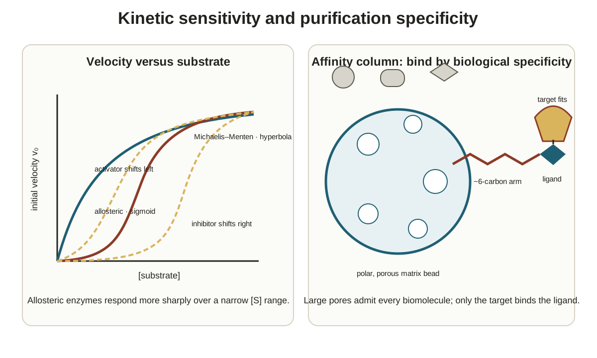
Initial velocity is zero when substrate concentration is zero, rises rapidly at low substrate, and levels as the enzyme becomes saturated or maxed out.
This hyperbolic curve is Michaelis–Menten behavior.
Most glycolytic enzymes, including LDH, were said to behave this way.
Calculating initial velocity V₀ at a chosen point requires substrate concentration [S], Vmax, and Km in the Michaelis–Menten equation.
Allosteric behavior
An allosteric enzyme gives a sigmoidal or S-shaped velocity curve.
Its related equation raises the substrate term to an exponent n.
n measures cooperativity among subunits.
Allosteric enzymes almost always have communicating subunits, and their communication uses weak bonds.
A larger n, such as 2.3 rather than 1.8, gives a more sharply rising curve.
Some glycolytic enzymes are allosteric.
Across the same bounded substrate-concentration range, Michaelis–Menten velocity changes little while allosteric velocity may rise or fall substantially.
An allosteric inhibitor shifts the sigmoidal curve to the right; the shift depends on inhibitor concentration.
An allosteric activator shifts the curve to the left.
Allosteric enzymes are sensitive to substrate, inhibitor, and activator concentrations in cells and experimental vessels.
Allosteric enzymes as metabolic valves
Cells use controlled pathway valves to direct intermediates according to current metabolic demands.
Glycolysis converts glucose to glucose 6-phosphate, but not every glucose 6-phosphate continues through glycolysis.
Some enters another pathway, such as the pentose phosphate pathway.
Dr. Hilt compared allosteric enzymes to valves that divert material according to cellular needs.
Reactions with highly negative ΔG values especially require tight control.
Research usually finds their enzymes to be allosteric, although Dr. Hilt noted that other control mechanisms also exist.
Affinity chromatography begins at the lecture boundary
Affinity chromatography isolates a particular biomolecule by exploiting biological specificity even when unwanted proteins have similar molecular weight or charge.
Figure · slide-derivedKinetic curves visualize enzyme sensitivity, while the affinity matrix captures one target through biological specificity rather than size exclusion Derived from annotated slide decks 1 and 2, pp. 19–20
Purification is necessary because cells are complex.
Dr. Hilt called an affinity column the current best method for isolating a particular biomolecule from a cell extract.
The column matrix begins as microscopic, flexible, cross-linked sugar-polymer beads.
Pores allow buffer and molecules to pass around and through a bead.
Something will be covalently attached to the bead, but its identity and construction were not completed in this lecture.
For enzyme purification, the bead should be polar.
Most surface residues of the example enzyme are polar, while most interior residues are nonpolar.
A nonpolar bead could cause the weak-bond-held protein structure to open or unfold, destroying enzyme activity.
The lecture stopped at this point, and the topic was to resume the next day.
Study Review Questions
Why do pH, weak bonds, enzymes, and energy form the course's initial foundation for studying intermediary metabolism?
Why do enzymes that catalyze reactions with very large negative ΔG values require especially strong control?
How does an animal cell function as an open chemical engine, and what do ΔG, ΔH, T, and ΔS describe in that framing?
What does the prime in ΔG′ specify, and how do the stated values for glucose breakdown and ATP synthesis compare?
How do DNA microarrays, two-dimensional gels, and microcalorimetry provide complementary views of cell metabolism?
How do mitochondria and chloroplasts organize the energy-conversion processes previewed for the last third of the course?
How does the LDH reaction connect hydride transfer, stereospecificity, proton consumption, buffer action, and glycolysis?
How would you reconstruct the calculation for making 1 L of 50 mM phosphate buffer at pH 6.8 from the stated stock and titrant?
How do Michaelis–Menten and allosteric velocity curves differ in shape, sensitivity, cooperativity, and response to inhibitors or activators?
Why must an affinity matrix for enzyme purification be polar, and what part of the column construction remained unfinished?