Included lectures:
| Label | Date | Source note |
|---|---|---|
| L1 | 2026-08-03 | notes/course-intro-metabolism-overview-2026-08-03.md |
| L2 | 2026-08-04 | notes/dna-microarrays-2d-gel-electrophoresis-2026-08-04.md |
| L3 | 2026-08-05 | notes/microcalorimetry-and-glycolysis-intro-2026-08-05.md |
| L4 | 2026-08-10 | notes/glycolysis-energetics-glycogen-2026-08-10.md |
Abbreviations
| Abbrev. | Meaning |
|---|---|
| G6P | Glucose-6-phosphate |
| F6P | Fructose-6-phosphate |
| F1,6BP | Fructose-1,6-bisphosphate |
| DAP | Dihydroxyacetone phosphate |
| G3P | Glyceraldehyde-3-phosphate |
| 1,3-BPG | 1,3-Bisphosphoglycerate |
| 3-PGA | 3-Phosphoglycerate |
| 2-PGA | 2-Phosphoglycerate |
| PEP | Phosphoenolpyruvate |
| HK | Hexokinase |
| PGI | Phosphoglucose isomerase |
| PFK1 | Phosphofructokinase-1 |
| TPI | Triosephosphate isomerase |
| G3PDH | Glyceraldehyde-3-phosphate dehydrogenase |
| PK | Pyruvate kinase |
| LDH | Lactate dehydrogenase |
| IEF | Isoelectric focusing |
| SDS | Sodium dodecyl sulfate |
| PAGE | Polyacrylamide gel electrophoresis |
| PI | Isoelectric point |
| IU | International unit |
| Pi | Inorganic phosphate |
| TPP | Thiamine pyrophosphate |
Prerequisites (BIS 102)
- Amino acid structures and one-letter codes (used without reteaching)
- Four levels of protein structure (primary through quaternary)
- Basic buffer chemistry and titration curves
- Spectrophotometry fundamentals
L1 -- Course Introduction and Metabolism Overview (2026-08-03)
Exam Structure and Grading
- Two midterms (50 min each) + one final (1 hr 50 min; most finish in ~1 hr)
- Final = 200 pts (2x midterm value); half covers post-MT2 material, half covers MT1+MT2
- Grading curve: class average = lowest B-; top ~15% receive some form of A
- Question types: calculation, short answer, some multiple choice (no Scantron); bring a calculator; all equations/constants furnished
- Exams test on lecture content and homework
EXAM: "You're crazy if you don't do the homework." (L1)
- HW1 is the only assignment for the first midterm (L1, reiterated L2)
Course Themes
| Theme | Key points |
|---|---|
| Weak bonds | Four non-covalent bond types; govern enzyme-substrate recognition, protein structure, membranes, dsDNA |
| Entropy (ΔS) | Important throughout; glucose (ordered) --> 6 CO2 (disordered) |
| Metal ions | Strict specificity at active sites (e.g., Mg²⁺ will not substitute for Mn²⁺ or Cu²⁺); some enzymes position two different metals |
| Proton gradients | ETC in mitochondria + photosystems 1 & 2 in chloroplasts; drives most ATP synthesis on the planet |
The Cell as a Chemical Engine
- Open system: nutrients in (glucose, O2), waste out (CO2, H2O, heat/entropy)
- No cell captures all consumed energy; some is always lost as heat and entropy
- ΔG = free energy for work; ΔH = enthalpy; T = temp (K); ΔS = entropy
- ΔG°′ for glucose --> CO2 + H2O = -686 kcal/mol
- ΔG°′ for ATP synthesis (ADP + Pi --> ATP) = +7.2 kcal/mol
- Pi structure at ~pH 7: P double-bonded to one O, two O- groups, one OH
- Course uses kcal/mol (Segel textbook); multiply by a constant to convert to joules
Three Techniques for Overall Cellular Metabolism (Overview)
| Technique | What it measures | Full coverage |
|---|---|---|
| DNA microarrays | Transcriptome (all mRNAs) | See L2 |
| 2D IEF-SDS PAGE | Proteome (all proteins) | See L2 |
| Microcalorimetry | Heat rate, CO2 production, O2 consumption | See L3 |
- Using multiple techniques provides different perspectives; relying on one method can be misleading (L1)
Mitochondria and Chloroplast Preview
- Mitochondria: PDH complex, Krebs cycle, fatty acid breakdown (matrix); ETC + ATP synthase (inner membrane); bacteria have ATP synthases in their inner membranes
- Chloroplasts: Photosystems 1 & 2 in thylakoid membranes (light reactions); Calvin cycle enzymes in stroma (CO2 --> sugar)
Buffers and Henderson-Hasselbalch
- Effective buffer requires: (1) high molarity (~50 mM+), (2) pKa near target pH, (3) appropriate B:A ratio (more B if reaction produces H+), (4) non-interfering with enzyme/detection
- Henderson-Hasselbalch: pH = pKa + log([B]/[A])
- At pH = pKa: 50% B, 50% A
- pH well above pKa: mostly base form
- When H+ added: [B] decreases, [A] increases by amount added; excess acid overwhelms buffer
- When H+ removed (e.g., consumed in a reaction): pH rises; A donates H+ to compensate
Buffer Preparation Problem: Phosphate Buffer at pH 6.8
- Goal: 1 L of 50 mM phosphate buffer at pH 6.8 from 1 M phosphate stock at pH 12, 2 M HCl, 3 M NaOH
- Volume of stock: 50 mmol / 1 M = 50 mL
- H-H at pH 12 (near pKa3 = 12.32): B (PO4 3-) = 32% (pH below pKa3, so < 50% fully deprotonated)
- H-H at pH 6.8 (near pKa2 = 7.21): A (H2PO4-) = 72% (pH below pKa2, so > 50% acid form)
- Total proton equivalents: 1.04 per mol phosphate --> 50 mmol x 1.04 = 52 mmol H+
- Volume HCl: 52 mmol / 2 M = 26 mL (NaOH not used -- pH must come down)
- Water: 1000 - 50 - 26 = 924 mL
- Shortcut: divide H-H ratio by 1, giving B/(B+A) directly without simultaneous equations
Man-Made Buffers: Tris
- Phosphate is in many biomolecules (DNA, RNA, phospholipids) and can interfere with enzymes; man-made buffers avoid this
- Tris structure: three CH2OH groups + one amino group on central carbon; CH2OH groups H-bond with water (instantly soluble)
- Single pKa (~8.2): acid form (protonated NH3+) = +1 charge; base form (lone pair on N) = 0 charge
- Preparation: start with tris base, add HCl to desired pH
Pyruvate-to-Lactate Reaction and NADH
Pyruvate + NADH + H+ --[LDH]--> L-Lactate + NAD+ (reversible; last step of glycolysis)- Three substrates, two products
- NADH carries a hydride (H-): two electrons + one proton
- Three oxidation states of H: H+ (no e-), H-dot (one e-), H- (two e-)
- NADH is a safe carrier: does not release energy upon bumping into membranes/proteins/DNA; only unlocked at enzyme active sites
- Pyruvate: 3 carbons; C1 carboxyl pKa 2.5 --> fully deprotonated at pH 7 (hence "pyruvate," not "pyruvic acid")
- LDH always produces L-lactate (OH on C2 drawn left); enzymes are stereospecific
- When reaction proceeds pyruvate --> lactate, a proton is consumed, which would raise pH without buffer
EXAM: Glycolysis must be known thoroughly for the first midterm -- structures, enzyme names, and whether each reaction is reversible. (L1)
Enzyme Kinetics: Michaelis-Menten vs. Allosteric
- Kinetics = how fast; thermodynamics = potential. A reaction can have large negative ΔG but without an enzyme has no chance kinetically.
| Feature | Michaelis-Menten | Allosteric |
|---|---|---|
| v0 vs. [S] curve | Hyperbola | Sigmoidal (S-shaped) |
| Equation | v0 = Vmax[S] / (Km + [S]) | v0 = Vmax[S]^n / (K0.5^n + [S]^n) |
| Subunits | Typically single | Multiple, cooperative (n = cooperativity coefficient) |
| Sensitivity | Less sensitive to [S] changes | Much more sensitive over same [S] range |
| Inhibitor effect | -- | Shifts curve right |
| Activator effect | -- | Shifts curve left |
| Role in regulation | -- | Act as valves controlling metabolite flow between pathways |
- Larger n --> steeper sigmoidal curve
- Reactions with large negative ΔG need tight control; usually catalyzed by allosteric enzymes
- Most glycolytic enzymes are Michaelis-Menten; a couple are allosteric (L1); specifically HK, PFK1, PK (L4)
- G6P can enter pentose phosphate pathway instead of continuing glycolysis; allosteric enzymes control which route (L1)
Affinity Chromatography (Introduction)
- Purpose: purify a specific biomolecule using biological specificity
- Column beads: microscopic, polar, flexible, cross-linked sugar polymer, porous
- Bead must be polar because most surface residues of a folded enzyme are polar; a nonpolar bead would unfold the enzyme
- See L2 for full affinity chromatography coverage
Key Definitions (L1)
| Term | Definition |
|---|---|
| Intermediary metabolism | Biochemical reactions in living cells |
| Assay | Measuring product made per unit time |
| ΔG°′ | Standard free energy change at 1 M concentrations, pH 7 |
| Vmax | Maximum enzyme velocity when saturated |
| Km | Michaelis constant; describes how well enzyme binds substrate |
| Turnover number | Substrates converted to product per enzyme molecule per minute (~30,000 for most enzymes) |
| KI | Equilibrium dissociation constant of an inhibitor |
| Transcriptome | Complete set of mRNAs a cell produces |
| Proteome | Complete set of proteins a cell produces |
L2 -- DNA Microarrays, 2D Gel Electrophoresis, and Lipid Review (2026-08-04)
Affinity Chromatography (Full Treatment)
- Beads are porous; the column is not performing molecular sieving or separating by size
- A covalent arm (~6 carbons recommended) lifts the ligand from the bead surface
- Too short (1-2 C): arm does not function
- Too long (12+ C): arm acts as hydrophobic coating
- Ligand: covalently attached to arm; can be substrate, product, inhibitor, coenzyme, or cofactor (metal ion)
- For enzymes with two substrates (e.g., HK uses glucose + ATP): only one substrate on the column; providing both would allow catalysis
- Enzyme binds ligand at active site via weak bonds (ionic, H-bond, van der Waals, hydrophobic effect); binding is dynamic but many ligands per bead retain the enzyme
- All other molecules flow through
Analytical Gel Electrophoresis
| Gel type | Separates by | Structure preserved | Notes |
|---|---|---|---|
| IEF | PI | All levels | Gentle; proteins migrate to PI and stop; current drops to ~0 when done; no tracking dye needed |
| Native PAGE | Size + charge-to-mass ratio (Q/M) | All levels | Cannot determine from one run whether faster band = smaller protein or higher Q/M |
| SDS-PAGE | Size only | Primary only | SDS denatures; gives all proteins same Q/M; mercaptoethanol breaks disulfide bonds |
- Stacking gel (~4%): compresses sample into thin band before entering resolving gel (~10%); prevents band spreading from diffusion (entropy)
- Wells formed by Teflon comb (~15 uL capacity each)
- Stacking requires different pH values in stacking gel, resolving gel, and upper buffer (resolving gel pH 8.8, stacking gel pH 6.8)
- IEF gels do not need stacking because proteins focus to sharp bands at PI as long as power is applied
- Bromophenol blue: tracking dye; does not interact with proteins; runs faster than any protein; signals when to stop
- SDS: 12-carbon man-made strong detergent (full -1 charge)
- Strong detergent = full charge; mild detergent = polar groups only
- Hydrophobic tail binds hydrophobic R groups (Leu, Ile, Trp, etc.); does not bind charged/polar residues (Glu, Arg) -- leaves gaps
- Larger subunit binds proportionally more SDS; thousands of negative charges overwhelm native charge --> same Q/M for all proteins
- SDS-coated denatured subunits repel each other --> clustered micelles
- Mercaptoethanol added in excess to reduce disulfide bonds (atmospheric O2 can create artificial disulfides)
EXAM: Correct electrode placement for SDS-PAGE is a likely MT1 question. SDS-coated proteins are heavily negative --> positive electrode at bottom. Negative electrode at bottom would drive proteins up into buffer. (L2)
Fatty Acids
| Name | Notation | Saturation | Double bond positions |
|---|---|---|---|
| Palmitic acid / palmitate | 16:0 | Saturated | -- |
| Stearic acid / stearate | 18:0 | Saturated | -- |
| Oleic acid / oleate | 18:1(9) | Unsaturated | C9-C10, cis |
| Linoleic acid / linoleate | 18:2(9,12) | Unsaturated | C9, C12, both cis |
| Alpha-linolenic acid / alpha-linolenate | 18:3(9,12,15) | Unsaturated | C9, C12, C15, all cis |
- Carbon 1 = carboxyl carbon; first number = total carbons; after colon = number of double bonds; parenthetical = positions
- All double bonds are cis (enzymes are stereospecific); cis creates a kink affecting membrane fluidity
- Ionized (COO-) form changes the name (e.g., palmitic acid --> palmitate)
- Free fatty acids act as detergents (compare SDS: 12C + charge vs. palmitate: 16C + charge); only sick/dying cells have lots of free fatty acids; normally covalently attached
- Stearic acid (18C) contains far more energy per molecule than glucose (6C)
EXAM: Students are typically asked to number the carbons in fatty acid structures. (L2)
Lipids: Acylglycerols
- Glycerol backbone with fatty acids at C1, C2, C3 via ester bonds
- Triacylglycerol (three fatty acids) = fat; energy deposit, not in membranes
- L-glycerol: OH on C2 drawn left; most organisms use L-form; archaea use D-glycerol
- Unique enzyme for each glycerol position (attach and remove); enzymes distinguish L- from D- using ~20 weak bonds
Lipids: Phospholipids
- Membrane construction (plasma membrane and organelle membranes)
- Structure: glycerol + fatty acid at C1 + fatty acid at C2 + phosphate at C3 linked to an alcohol via phosphodiester
- Phosphodiesters also in DNA/RNA backbones
| Phospholipid | Head group | Charge note |
|---|---|---|
| Phosphatidylcholine | Choline (quaternary amine: N with 4 groups, always +1 regardless of pH) | |
| Phosphatidylserine | Serine (amino acid; functions beyond protein building) |
- Phosphate is always at C3; the enzyme is stereospecific and never makes a mistake (mutation would likely be lethal)
EXAM: Students may be asked to draw a specific phospholipid at a given pH with designated fatty acids at each position. (L2)
DNA Microarrays (Full Procedure)
mRNA isolation:
- Extract all RNA with organic solvent; only mRNA is wanted
- mRNA distinguished by poly-A tail at 3' end (tRNA, rRNA lack it)
- Affinity column with oligo-dT ligand (covalently attached directly to bead; no arm needed -- mRNA is linear)
- mRNA poly-A tails bind oligo-dT via H-bonding (A-T); dynamic binding; abundance of oligo-dT retains mRNA
- tRNA and rRNA flow through
- Two identical columns: one for cancer cells, one for normal cells (control)
- Fritted disc at bottom traps beads
- Nucleic acids absorb UV at 260 nm (A260; conjugated double bonds in bases)
- Flow-through: high A260 fractions = tRNA + rRNA (discard); wash with buffer (~pH 7.5) until A260 = 0
Elution:
- Change pH by large amount (disrupts H-bonds) or decrease salt concentration
- Decreasing salt removes cation shielding from phosphate backbone negative charges --> strands repel and separate
cDNA synthesis and labeling:
- Add excess oligo-dT primers (anneal to poly-A tail for double-stranded starting point)
- Add reverse transcriptase
- Add dNTPs (dATP, dGTP, dCTP, dTTP) -- energy sources and building blocks
- One dNTP (e.g., dATP) made fluorescent (emits at longer wavelength when excited)
- Reverse transcriptase synthesizes cDNA from mRNA template
- Cancer cell mRNA --> red fluorescent cDNA; normal cell mRNA --> green fluorescent cDNA
- Two outcomes: mRNA gains visible color; copy is DNA (more stable than RNA)
Removing mRNA template:
- Incubate mRNA-cDNA duplex in 1 M NaOH + heat for 15 min
- Mechanism: OH- deprotonates 2'-OH of RNA sugar --> 2'-O- attacks partial-positive phosphorus --> breaks phosphodiester bond at every nucleotide --> fragments mRNA completely
- DNA is unaffected (no 2'-OH; has only H at 2' position)
- Critical: add HCl to neutralize back to ~pH 7 (cDNA cannot H-bond to chip at high pH)
The chip:
- Microscope slide; each dot = multiple copies of one gene's ssDNA (~1 nL volumes, machine-deposited)
- Adjacent dots = different genes; each dot must have ~same copy number
- ssDNAs act as receptors; gene identity at each position known only to manufacturer
Hybridization and scanning:
- Equal amounts red + green cDNA incubated with chip + buffer (optimized); rotated
- Rinse to remove non-specific binding; place in fluorescence scanner
| Spot color | Interpretation |
|---|---|
| Red | Gene more expressed in cancer cells |
| Green | Gene more expressed in normal cells |
| Yellow | Equal expression in both |
| Black | Gene not expressed in either |
- Scanner images red and green channels separately, then merges
- Red spots identify genes with altered expression in cancer; not all may be direct causes (one upstream gene could control many targets)
2D IEF-SDS PAGE (Full Procedure)
Advantage over microarrays: A single base substitution changing a residue's charge (e.g., Val --> Glu) shifts the protein on IEF but may not be detected by a microarray (rest of mRNA still binds). SDS-PAGE alone will not detect charge changes.
First dimension -- IEF tube gel:
- Glass tube, gel polymerizes in ~15 min
- Components: acrylamide (monomer), bisacrylamide (cross-linker; without it no gel forms), ampholytes (small molecules creating pH gradient when current applied), TEMED (prolongs free radical lifetime), ammonium persulfate (persulfate bond breaks in water --> sulfate free radicals --> initiate polymerization chain reaction)
- Electrodes: top = 60 mM NaOH (high pH), bottom = 60 mM H2SO4 (low pH); extreme pH traps ampholytes in gel
- Broad pH gradient (pH 2-10) for all proteins; sample loaded on top; proteins migrate to PI and stop
- Hundreds of bands possible; some proteins may co-migrate
SDS treatment of extruded IEF gel:
- Gel extruded from tube with plunger (like spaghetti) into SDS solution
- SDS diffuses into gel; proteins too large to diffuse out
- SDS + mercaptoethanol + heat: separates subunits, completely unfolds (only primary structure remains)
Second dimension -- SDS-PAGE slab gel:
- Single large well (Teflon comb) to accommodate entire IEF gel
- Pour resolving gel first, then stacking gel; place IEF gel in well
- Stacking ~4%, resolving ~10%; SDS throughout
- Positive electrode at bottom (SDS-coated proteins heavily negative, migrate down)
- Bromophenol blue + glycerol on top for tracking; stop when dye nears bottom
- Result: 2D map; vertical line = same PI; horizontal line = same MW; smaller proteins toward bottom
- Stain to visualize; merge cancer vs. normal images to find differences
- Sensitivity enhancement: grow cells on S-35 methionine + autoradiography (~1 week in freezer)
L3 -- Microcalorimetry and Introduction to Glycolysis (2026-08-05)
Microcalorimetry (Full Treatment)
- Third technique for examining cells; measures very small heat flows (microwatt range) from living cells
- Differs from freshman chem calorimetry: cells stay alive, not burned to completion
- Can also measure CO2 production rate and O2 consumption rate
- These three measurements cannot be obtained from microarrays or 2D gels (L3)
- ΔG = ΔH - T ΔS; microcalorimeter measures only ΔH
EXAM: The Gibbs free energy equation is always provided on exams. (L3)
Instrument setup:
- Ampule: small metal container (~1 mL), special heat-conducting alloy; welded metal cylinder inside creates an upper well (~40 uL)
- Tissue (leaves, bacteria, RBCs, etc.) in main chamber; must not contact liquid in upper well
- Metal lid screws on; small hole fitted with thin tubing to pressure sensor (thin to prevent external heat conduction)
- Instrument: 4 openings -- 1 reference (empty, never changed), 3 sample positions; each surrounded in 3D by heat sensors --> electrical signals --> computer (separate channels)
- Large lid covers entire instrument to block room heat
Part A -- Baseline heat rate (~30 min):
- Upper well: 40 uL water
- y-axis: microwatts/mg dry weight; x-axis: time (min)
- Initial signal off-scale (room heat); recovers and levels off at plateau = heat flow from living cells
- If signal = 0, cells are dead
Part B -- CO2 trap (~30 min):
- Replace water with 40 uL of 0.4 M NaOH (strong base, deliberately in excess); NaOH never touches tissue
- Signal levels off at higher plateau than Part A
- Difference (B - A) is proportional to CO2 production rate
- Mechanism: OH- attacks partial-positive C of CO2 --> bicarbonate; excess OH- converts to carbonate; reactions are exothermic
- CO2 is nonpolar, crosses membranes freely; in sealed ampule, bounces at high velocity (ideal gas); hitting NaOH surface = chemically trapped as carbonate + heat
- NaOH in excess ensures every CO2 captured
- If Part B = Part A, no detectable CO2 (nonfunctional mitochondria in plant cells)
Part C -- Control (~30 min):
- Replace NaOH with 40 uL water
- Signal should return to ~Part A baseline
- Verifies Part B caused no tissue damage (e.g., NaOH dripping onto tissue)
Why water in Part A: Controls for water vapor; NaOH solution is mostly water. Ensures Part B effect is due to NaOH, not water vapor changing cell metabolism.
Dry weight determination: After experiment (~90 min total), tissue dried in vacuum oven at 90 C overnight (vacuum prevents oxidation); weighed on precise balance.
Pressure sensor and O2 measurement:
- Pressure data collected simultaneously throughout
- Parts A and C: multiple gases (N2, O2, CO2) --> cannot separate individual contributions
- Part B: NaOH eliminates CO2, N2 is inert --> any pressure decrease = O2 consumption (mainly mitochondria)
- Calculate picomoles O2/min using PV = nRT
Summary of output: Heat rate/mg dry weight, CO2 production rate, O2 consumption rate (all with real numerical values)
Application: Predicts tree growth potential in 2-3 days from small tissue samples (vs. years of field observation); also applicable to cancer cells; many replicates (~20) for averaging
Intermediary Metabolism
- Studies small organic compounds (~1,000 MW or less): ATP, glucose, fatty acids, amino acids -- not macromolecules
- Catabolic reactions break down compounds; converge on acetyl-CoA
- Anabolic reactions build compounds; diverge/branch from common precursors
- Energy carriers connecting the two: NAD/NADH, NADP/NADPH, FAD/FADH2, ATP
- These exist in small pool sizes; cells do not store large quantities
- To build something (needs ATP, etc.), cell must simultaneously break something down; pathways are perfectly meshed
Pathway types:
| Type | Description | Example |
|---|---|---|
| Convergent (catabolic) | Many substrates funnel into one compound | Glucose, glycogen, fatty acids --> acetyl-CoA |
| Divergent (anabolic) | Pathways branch for biosynthesis | -- |
| Cyclic | Intermediates recycled; only tiny amounts needed | Krebs cycle: 2C in as acetyl-CoA, 2C out as CO2 |
- Acetyl-CoA: 2-carbon acetyl group carried by CoA via thioester (S-C=O)
EXAM: Students must know glycolysis and the Krebs cycle thoroughly. (L3)
Coupled Enzyme Reactions: Glutamine Synthetase
NH4+ at high concentration is toxic; many N-containing compounds break down to ammonia --> ammonium at cell pH
Glutamate + NH4+ --> glutamine is endergonic alone
Cells couple it with exergonic ATP breakdown at one enzyme active site:
Glutamate + NH4+ + ATP --[Glutamine synthetase]--> Glutamine + ADP + PiMechanism:
- ATP phosphorylates side-chain carboxyl of glutamate (never the alpha-carboxyl; enzyme orients correctly) --> acyl phosphate intermediate + ADP
- NH4+ enters active site; active-site group removes H+ --> NH3; nitrogen lone pair (nucleophile) attacks electrophilic carbonyl C of acyl phosphate --> displaces phosphate --> glutamine + Pi
EXAM: Acyl phosphates are high-energy compounds (large negative ΔG); recognize them immediately. (L3)
- Coupled reactions were discovered early through glycolysis study; led to incorrect assumption that all energy-transfer reactions use substrate-level coupling (proton gradients discovered later)
- Coupled reactions need not involve ATP
Glycolysis Steps 1-4
Glycolysis = lysis of sugars; occurs in cytosol; glucose --> pyruvate/lactate. Continues in L4.
Glucose structure:
- Beta-D-glucose: 6 carbons; C1 at anomeric position
- Shorthand: vertical lines = OH groups; C2 OH below, C3 OH above, C4 OH below
- C1 OH above = beta (slightly more stable in solution); below = alpha; ring opens/closes (mutarotation)
Glucose transport:
- Glucose is very polar (multiple O atoms); cannot cross nonpolar membrane interior without help
- Glucose transporter: 45,000 MW membrane protein; facilitates diffusion both directions
- Humans have 14 forms (GLUT1-14); brain runs mostly on glucose
Step 1: Hexokinase
Glucose + ATP --[HK; Mg²⁺]--> G6P + ADP
- Phosphorylates C6 OH specifically; "hexokinase" implies it handles other hexoses too
- At physiological pH, phosphate on C6 carries -2 charge
- Consumes 1 ATP (even though glycolysis goal is to produce ATP)
- Accomplishments: (1) pulls equilibrium (glucose disappears, more enters cell), (2) traps glucose inside (transporter rejects G6P), (3) charged handle on C6 helps next enzyme orient substrate, (4) raises glucose energy slightly
- Mg²⁺ required: sits near ATP phosphates, shields negative charges so C6 OH (nucleophile, partial negative) can approach and attack phosphorus (partial positive). Without Mg²⁺, no attack.
- HK conformational change: two domains; open without glucose; glucose binding closes domains like a clamshell, burying glucose and physically excluding water. C6 OH attack is 40,000x faster than water attack. (L3)
Step 2: Phosphoglucose Isomerase (PGI)
G6P --[PGI]--> F6P (reversible)
- Moves bond from ring O to C2; converts C1 from aldehyde to alcohol (aldose --> ketose)
- Purpose: aldehyde C1 is electrophilic (not a nucleophile); converting to OH allows C1 to attack ATP in step 3
Step 3: Phosphofructokinase-1 (PFK1)
F6P + ATP --[PFK1]--> F1,6BP + ADP (one-way, irreversible)
- Phosphorylates C1 OH using a second ATP; glycolysis has now consumed 2 ATPs
- A second isomer (PFK2) exists; discussed later in course
Step 4: Aldolase
F1,6BP --[Aldolase]--> DAP + G3P (reversible)
- Splits F1,6BP between C3 and C4 --> two 3C molecules, each with a phosphate
- Reverse = aldol condensation (L4)
- Cell strategy: phosphorylate C6, phosphorylate C1, split down the middle
L4 -- Glycolysis Completion, Energetics, and Beer's Law (2026-08-10)
Covers sections 1-21 of the source note only. The lecturer stated in section 22: "Material from this point forward is on the second midterm." Post-boundary content (starch, glycogen) is outside this guide's scope.
Glycolysis Steps 5-10
Step 5: Triosephosphate Isomerase (TPI)
DAP --[TPI]--> G3P
- Converts DAP to G3P; glucose effectively yields 2 G3P molecules
- TPI is catalytically perfect: cannot evolve faster; only rate limitation is substrate diffusion
- Without TPI, half of glucose's carbons would be lost
Step 6: Glyceraldehyde-3-Phosphate Dehydrogenase (G3PDH)
G3P + Pi + NAD+ --[G3PDH]--> 1,3-BPG + NADH + H+ (x2 per glucose)
- Dehydrogenases transfer electrons onto NAD, FMN, or FAD; kinases transfer phosphate
- 1,3-BPG contains a high-energy acyl phosphate bond (ΔG°′ ~ -12 kcal/mol)
- If water were used instead of Pi, energy would be lost as heat/entropy; Pi traps energy in acyl phosphate for ATP synthesis in next step
- H+ released comes from Pi (has OH at physiological pH)
- NAD+ reduced by hydride (H-): H and its two bonding electrons transfer from substrate C to NAD
- 1,3-BPG is the cousin of 2,3-BPG (important for hemoglobin)
Step 7: Phosphoglycerate Kinase
1,3-BPG + ADP --[Phosphoglycerate kinase]--> 3-PGA + ATP (reversible; x2 per glucose)
- 3-PGA: C1 = carboxyl group, phosphate on C3
- Net ATP at this point from 1 glucose: 2 consumed (HK + PFK1), 2 produced here --> net = 0 (break even)
Step 8: Phosphoglycerate Mutase
3-PGA --[Phosphoglycerate mutase]--> 2-PGA
- 3-PGA and 2-PGA are structural isomers
- Shell game mechanism: active site already holds a phosphate; places it on C2 before removing the one on C3 (the phosphate on C2 is not the same one that was on C3)
- Direct removal + reattachment would cost too much energy
Step 9: Enolase
2-PGA --[Enolase; removes H2O]--> PEP
- Removes OH on C3 + H on C2 as water --> double bond with phosphate on C2
- PEP ΔG°′ = -13.8 kcal/mol (more than enough to make an ATP)
- PEP's instability: without the phosphate, molecule would tautomerize enol --> keto (more stable); phosphate pins it, preventing tautomerization until next enzyme
Step 10: Pyruvate Kinase
PEP + ADP --[PK]--> Pyruvate + ATP (one-way; x2 per glucose)
- Phosphate released and donated to ADP; keto-enol tautomerization proceeds --> large negative ΔG
LDH and NAD Regeneration
Pyruvate + NADH + H+ --[LDH]--> Lactate + NAD+
- Purpose: regenerate NAD+ (pool size is very small); without this step, G3PDH reaction stalls and glycolysis stops
- Net yield from 1 glucose: 2 ATP
EXAM: Students need to know the steps of glycolysis thoroughly. (L4)
Complete Glycolysis Summary Table
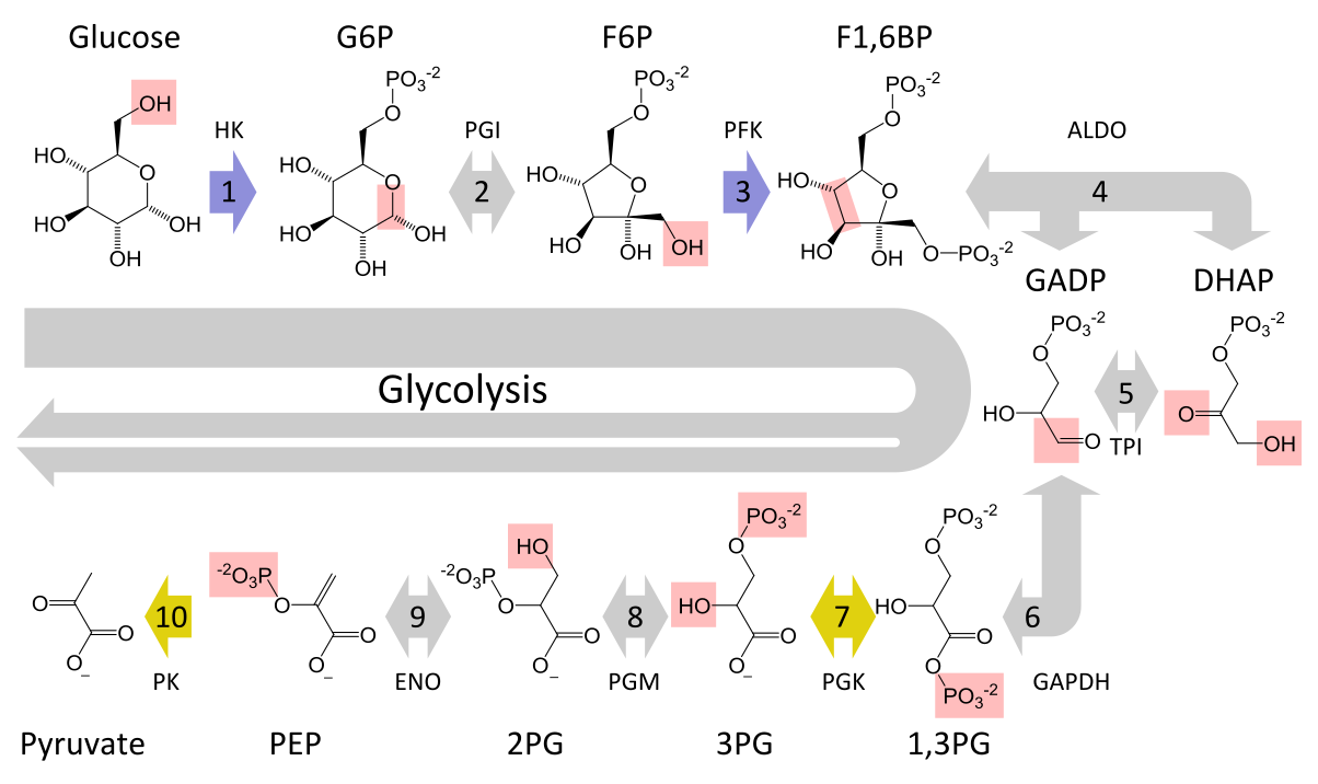

| Step | Enzyme | Reaction | Reversible? | ATP | Notes |
|---|---|---|---|---|---|
| 1 | Hexokinase | Glucose + ATP --> G6P + ADP | No | -1 | Mg²⁺ required; allosteric |
| 2 | PGI | G6P --> F6P | Yes | -- | Aldose --> ketose |
| 3 | PFK1 | F6P + ATP --> F1,6BP + ADP | No | -1 | Allosteric |
| 4 | Aldolase | F1,6BP --> DAP + G3P | Yes | -- | Splits at C3-C4 |
| 5 | TPI | DAP --> G3P | Yes | -- | Catalytically perfect |
| 6 | G3PDH | G3P + Pi + NAD+ --> 1,3-BPG + NADH + H+ | Yes | -- | x2; acyl phosphate |
| 7 | Phosphoglycerate kinase | 1,3-BPG + ADP --> 3-PGA + ATP | Yes | +2 | x2; break-even point |
| 8 | Phosphoglycerate mutase | 3-PGA --> 2-PGA | Yes | -- | Shell game mechanism |
| 9 | Enolase | 2-PGA --> PEP + H2O | Yes | -- | PEP is high-energy |
| 10 | Pyruvate kinase | PEP + ADP --> Pyruvate + ATP | No | +2 | Allosteric |
| -- | LDH | Pyruvate + NADH + H+ --> Lactate + NAD+ | Yes | -- | Regenerates NAD+ |
Net: 2 ATP per glucose (both glycolysis and alcoholic fermentation)
Alcoholic Fermentation in Yeast
- Glucose processed through glycolysis to pyruvate; last two steps differ from lactate pathway:
Pyruvate --[Pyruvate decarboxylase; Mg²⁺ (cofactor), TPP (coenzyme)]--> Acetaldehyde + CO2 (one-way)- CO2 is nonpolar, leaves the cell
- Pyruvate decarboxylase is allosteric (one-way reaction)
Acetaldehyde --[Alcohol dehydrogenase]--> Ethanol + NAD+- Purpose: regenerate NAD+ (same logic as LDH -- small pool size)
- Net: 2 ATP per glucose
EXAM: If asked to draw all steps of alcoholic fermentation, include the entire glycolysis pathway, not just the last two steps. (L4)
NAD Structure and Spectral Properties
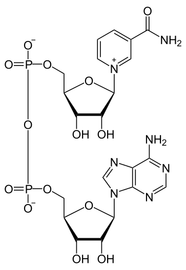

- NAD = nicotinamide adenine dinucleotide
- Lower portion = AMP (acts as handle for enzyme orientation; AMP handles also in CoA, FAD, etc.)
- Business end = nicotinamide ring (amide: C=O bonded to NH2, not amine)
- Hydride (H-) placed specifically at top of nicotinamide ring and nowhere else
- Positive charge on N of NAD+ partially delocalized by resonance to C at top of ring --> attracts hydride
- Reduction of NAD+ to NADH eliminates the positive charge on N
- Students should be able to draw both oxidized (NAD+) and reduced (NADH) forms
| Wavelength | NAD+ absorbs? | NADH absorbs? | Use |
|---|---|---|---|
| 340 nm | No | Yes | Measure NADH specifically; buffer, proteins, NAD+, substrates do not absorb here |
| 260 nm | Yes | Yes | Both absorb strongly with only a slight, unusable difference; proteins and nucleic acids also absorb strongly |
Allosteric Regulation of Glycolysis
- Generalization: reactions with large negative ΔG --> typically catalyzed by allosteric enzymes (multiple subunits, sigmoidal kinetics)
- Allosteric glycolytic enzymes: HK, PFK1, PK (all catalyze one-way reactions)
- In alcoholic fermentation: pyruvate decarboxylase is also allosteric (one-way)
- Allosteric enzymes are much more sensitive to changes in compound concentrations
- PFK1 regulation:
- ADP = positive allosteric effector (activator): ADP buildup --> cell low on energy --> PFK1 speeds up
- ATP = negative allosteric effector (inhibitor): ATP buildup --> cell has enough energy --> PFK1 slows down
- ADP binds at a site separate from active site, changes conformation, increases rate
- Nothing in metabolism ever goes to zero; negative regulation = rate slows down
Purposes of Glycolysis
- Produce some ATP (net 2 per glucose)
- Provide intermediates siphoned off for biosynthesis:
- 3-PGA --> serine (S) --> glycine or cysteine
- DAP --> glycerol (backbone of phospholipids and triacylglycerols)
- 1,3-BPG --> 2,3-BPG in red blood cells (for hemoglobin function; RBCs sacrifice glycolysis to make it)
Equilibrium and Free Energy
- K_eq = [products] / [substrates] at equilibrium
- Cells never reach equilibrium; operate in steady state (open system). Biochemistry uses equilibrium math as a simplification.
- K_eq′: K_eq measured at pH 7 (for reactions involving H+)
- ΔG = RT ln([products]/[substrates]) - RT ln(K_eq)
- At equilibrium: ΔG = 0; reaction cannot do work
- If all species at 1 M: ln(products/substrates) = ln(1) = 0 --> chosen as reference to simplify math
- ΔG° = -RT ln(K_eq)
- If all species at 1 M and pH 7: ΔG°′
- Actual cell free energy: ΔG = ΔG°′ + RT ln(actual product/substrate ratio)
- ΔG°′ values are reference values for comparing reactions in tables; real ΔG depends on actual concentrations (change throughout the day)
- Reversing a reaction: sign of ΔG reverses, K_eq inverts
- Adding (coupling) two reactions: ΔG values add, K_eq values multiply
EXAM: Equations will be provided; students need to understand what they mean. (L4)
Beer's Law
- A = epsilon x L x C
- A = absorbance (unitless); spectrophotometer measures I0 and I, calculates log10(I0/I)
- epsilon = molar extinction coefficient (M^-1 cm^-1); large number; determined by molecular structure; requires conjugated double bonds
- L = path length (cm); standard = 1 cm
- C = concentration (M, moles/liter); typically very small in biochemistry
- Equation of a straight line: A directly proportional to C, passes through zero
- Absorbance proportional to concentration, not mass; spectrophotometer does not integrate over volume
- Linearity must be verified experimentally; curves off at high concentrations
- Practical test: dilute sample in half, check that A is halved
- Alternative form: A = a x L x C, where little a = specific absorbance; used when MW unknown; C in mg/mL or g/100 mL. Cannot calculate IUs with this form (IUs require micromoles/min --> need MW).
LDH Enzyme Assay and IU Calculation
Reaction: Pyruvate + NADH + H+ --[LDH]--> Lactate + NAD+- At 340 nm: only NADH absorbs (pyruvate, H+, LDH protein, buffer do not)
- At t=0, A340 is high (NADH present); if LDH present, A340 decreases as NADH consumed; flat line = no LDH
- Use initial linear region (Beer's Law region); slope = initial velocity (v0)
- Epsilon for NADH at 340 nm: 6,220 M^-1 cm^-1 (would be given on exam)
IU calculation steps:
- Measure Delta A / time from linear region
- Convert to per-minute change (e.g., 15 s measurement x4)
- Calculate Delta [NADH] (M/min) using Beer's Law: Delta C = Delta A / (epsilon x L)
- Multiply by assay volume (L) to convert M --> moles (liters cancel)
- Convert moles --> micromoles (x 10^6)
- Result = IUs of active LDH in cuvette
- Example: 0.15 umol NADH consumed/min = 0.15 IU active LDH
- NADH change is negative (consumed), but IUs reported as positive
- Always carry units through calculations
EXAM: HW1 covers the IU calculation for MT1. HW2 and final extend it further (e.g., IUs in original extract). (L4)
Review Questions
In the microcalorimetry experiment, what specific chemical mechanism causes the Part B heat signal to exceed Part A, and what control verifies that the difference is genuinely due to CO2? (L3)
Explain why SDS-PAGE separates proteins by size only, while native PAGE separates by two parameters. What structural information is lost under SDS treatment? (L2)
Walk through the complete IU calculation for LDH: given a measured Delta A at 340 nm over a known time interval, epsilon = 6,220 M^-1 cm^-1, L = 1 cm, and a known assay volume, derive the IUs of active LDH. (L4)
Why does hexokinase require Mg²⁺ as a cofactor, and how does the enzyme's conformational change upon glucose binding explain why the C6 hydroxyl attacks 40,000 times faster than water? (L3)
PEP has a ΔG°′ of -13.8 kcal/mol. What structural feature makes PEP a high-energy compound, and how does pyruvate kinase exploit this? (L4)
In the DNA microarray procedure, why does incubation in 1 M NaOH destroy the mRNA template but leave the cDNA intact? (L2)
At what point in glycolysis does net ATP production reach zero (break even), and what is the final net ATP yield per glucose? Identify which enzymes consume and which produce ATP. (L3, L4)
How do allosteric activators and inhibitors of PFK1 (ADP and ATP, respectively) regulate glycolytic flux in response to the cell's energy status? (L4)
Draw or describe the structure of a phospholipid with palmitate at position 1 and oleate at position 2. Where does the phosphate attach, and why is this placement absolute? (L2)
Besides ATP production, what is the second major purpose of glycolysis? Name two intermediates that are siphoned off and what they are used to build. (L4)
Known-Gaps Appendix
- Post-boundary L4 content (second-midterm material): The lecturer stated in the Aug 10 lecture that material from starch/glycogen onward is on the second midterm. The following topics from the L4 source note are therefore outside this guide's scope: starch structure (amylose, amylopectin), alpha-1,4 and alpha-1,6 glycosidic bonds, reducing vs. non-reducing ends, glycogen structure and function, glycogenin, glycogen synthase, and UDP-glucose. These will be covered in a Midterm 2 study guide.